


 TR
 EN
 RU
 AR
 FR


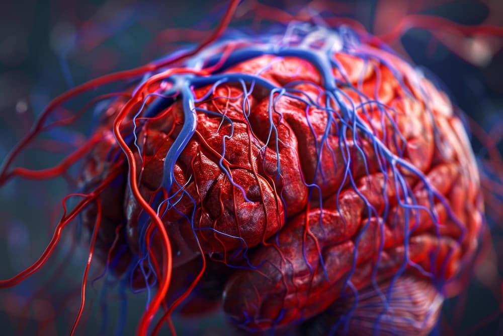


An arteriovenous malformation, often called an AVM, is an abnormal tangle of blood vessels that connects arteries directly to veins. Normally, tiny blood vessels called capillaries sit between arteries and veins to slow blood flow and deliver oxygen to tissues. In an AVM, blood bypasses this normal network, which can place extra pressure on the blood vessels.
A brain AVM may remain silent for years and be found incidentally during an MRI or CT scan performed for another reason. However, an AVM can rupture and cause bleeding in the brain, known as a hemorrhagic stroke. This guide explains the types of AVM, possible symptoms, how AVMs are diagnosed, their relationship with stroke, and available treatment options.
What Is an Arteriovenous Malformation?
An arteriovenous malformation is a type of vascular malformation. It involves an abnormal connection between arteries, which carry blood away from the heart, and veins, which return blood to the heart.
In a healthy circulatory system, blood flows from arteries into capillaries before moving into veins. Capillaries reduce the pressure of blood flow. In an AVM, high-pressure arterial blood flows directly into veins that are not designed to handle it. This can make the blood vessels enlarge, weaken, or bleed.
AVMs can occur in different areas of the body. A cerebral arteriovenous malformation, also called a brain arteriovenous malformation, affects blood vessels in the brain and is the most widely recognized type.
Congenital vs. Acquired AVM
Most arteriovenous malformations are congenital, meaning they develop before birth as the blood vessels are forming. In these cases, arteries and veins do not develop with their usual capillary network between them. Instead, high-pressure blood from an artery flows directly into nearby veins.
A congenital AVM may be present from birth but cause no symptoms for many years. Some people only learn they have an AVM after having an MRI or CT scan for a different reason. Others are diagnosed after symptoms such as headaches, seizures, weakness, or bleeding occur.
In most cases, a congenital AVM is not directly inherited and does not result from anything a parent did during pregnancy. It is not caused by diet, exercise, stress, childhood injuries, or daily activities. However, a small number of AVMs can occur in association with inherited conditions, such as hereditary hemorrhagic telangiectasia (HHT), which affects blood vessel development.
An acquired arteriovenous malformation develops later in life rather than before birth. It is much less common than a congenital AVM and may occur after an injury, surgery, infection, or another condition that damages blood vessels or changes how blood flows through an area.
Some acquired artery-to-vein connections are described more accurately as an arteriovenous fistula rather than an AVM. An arteriovenous fistula is usually a single direct connection between an artery and a vein, while a typical AVM contains a more complex tangle of abnormal blood vessels.
AVM vs. Aneurysm and Other Vascular Malformations
An AVM and an aneurysm are different conditions. An aneurysm is a weak, bulging area in the wall of an artery. An AVM is a cluster of abnormal vessels where arteries connect directly to veins.
A cavernous malformation is another type of blood vessel abnormality. It is made of small, tightly packed vessels with slower blood flow than an AVM. It may bleed, but its appearance and treatment approach can differ from those of a brain AVM.
An arteriovenous fistula is a direct connection between an artery and a vein. Unlike an AVM, it does not usually involve a tangled network of abnormal blood vessels. An arteriovenous fistula may be present at birth or develop later because of trauma, surgery, or other medical conditions.
Types of AVM by Location
An AVM can develop in different parts of the body. Symptoms, possible complications, and treatment options depend largely on its location, size, and the blood vessels involved.
Brain (Cerebral) AVM
A brain AVM is an abnormal connection between blood vessels in the brain. It may cause no symptoms, especially if it is small or located away from areas that control speech, movement, vision, or sensation.
However, a brain AVM can bleed into surrounding brain tissue. This is an emergency because bleeding can raise pressure inside the skull and damage brain cells. A ruptured brain AVM can lead to a hemorrhagic stroke.
Possible effects of a brain AVM include:
Headaches
Seizures
Weakness or numbness on one side of the body
Problems with balance or coordination
Speech or language difficulties
Changes in vision
Memory or concentration problems
Developmental concerns in children
Brain hemorrhage
Spinal AVM
A spinal AVM affects blood vessels in or around the spinal cord. It can interfere with blood flow to the spinal cord and may gradually affect movement, sensation, bladder control, or bowel control.
Symptoms can develop slowly and may be mistaken for other spinal conditions. In some cases, bleeding from a spinal AVM can cause sudden back pain, weakness, or paralysis.
Possible spinal AVM symptoms include:
Progressive weakness in the legs or arms
Numbness, tingling, or altered sensation
Difficulty walking
Loss of balance
Bladder or bowel changes
Sudden severe symptoms if bleeding occurs
Pulmonary AVM
A pulmonary arteriovenous malformation occurs in the lungs. It allows blood to pass from the right side of the circulation to the left side without receiving its usual supply of oxygen in the lungs.
Some people have no symptoms. Others may experience breathlessness, low oxygen levels, blue-colored lips or fingertips, tiredness, or migraine-like headaches. Pulmonary AVMs can also allow small clots or bacteria to bypass the lungs’ filtering system and reach the brain, increasing the risk of stroke or brain infection.
Pulmonary AVMs may occur in people with hereditary hemorrhagic telangiectasia, also known as HHT. HHT is an inherited condition that can cause fragile blood vessels and repeated nosebleeds.
Peripheral AVM
A peripheral AVM affects blood vessels outside the brain, spinal cord, and lungs. It may occur in the skin, face, arms, legs, pelvis, or other tissues.
A peripheral AVM may appear as a warm, reddish, bluish, or swollen area. It can enlarge over time and may cause pain, throbbing, skin changes, bleeding, or difficulty using the affected body part.
Possible signs include:
A visible or raised blood vessel area
Warmth over the skin
Pulsing or throbbing
Pain or tenderness
Swelling
Skin discoloration
Bleeding from the affected area
Reduced movement or function in a nearby joint or limb
Symptoms of AVM
AVM symptoms vary depending on the location and whether bleeding has occurred. Some people have a silent AVM, also called an asymptomatic AVM, and only learn about it after an imaging test for a different health concern.
An incidental AVM finding can be worrying, but it does not automatically mean emergency treatment is needed. A specialist team considers the AVM’s location, size, blood-flow pattern, past bleeding, symptoms, and treatment risks before making recommendations.
Signs of an Unruptured AVM
An unruptured AVM has not caused a major bleed. It may cause no symptoms, or symptoms may develop gradually because of changes in blood flow or irritation of nearby tissue.
Brain AVM symptoms may include:
Headaches, including recurring or unusual headaches
Seizures
Weakness, numbness, or tingling
Problems with speech or understanding language
Vision changes
Dizziness or balance problems
Difficulty concentrating
Facial pain or changes in sensation
Developmental delays or learning difficulties in some children
An AVM headache can have many possible causes, and most headaches are not caused by an AVM. However, new, severe, worsening, or unusual headaches should be discussed with a healthcare professional.
Signs of AVM Rupture
An AVM rupture can cause sudden bleeding, especially when the AVM is located in the brain. This is a medical emergency.
Call emergency services immediately if a person develops:
A sudden, severe headache unlike previous headaches
Loss of consciousness or fainting
A seizure
Sudden weakness or numbness, especially on one side
Facial drooping
Trouble speaking, understanding, or seeing
Severe dizziness, loss of balance, or difficulty walking
Sudden confusion
Repeated vomiting
Sudden, severe neck pain
These symptoms can be signs of an AVM hemorrhage or stroke. Rapid assessment is essential because urgent treatment may reduce the risk of long-term brain injury.
What Causes AVM and Who Is at Risk?
Most AVMs form during early development before birth. The exact reason why this happens is not fully understood. In most cases, there is no known action a parent or patient could have taken to prevent an AVM.
AVMs are usually sporadic, meaning they occur without a known family history. However, some AVMs may be associated with genetic conditions, particularly hereditary hemorrhagic telangiectasia.
Factors that may be relevant include:
Having HHT or a family history of HHT
A history of recurrent nosebleeds or visible small blood vessels on the lips, tongue, face, or hands
Previous trauma, surgery, or infection in cases of acquired arteriovenous malformation
A known vascular condition affecting blood vessels
An AVM can occur at any age. AVM in children may be detected because of seizures, headaches, developmental concerns, or bleeding. Some people are not diagnosed until adulthood, often after symptoms develop or an imaging test identifies the condition.
How Is AVM Diagnosed?
AVM diagnosis often begins after symptoms such as seizures, unusual headaches, or stroke-like signs. It may also begin after an incidental finding on imaging. The diagnostic process helps doctors identify the AVM’s location, size, shape, blood supply, and drainage pattern.
An AVM MRI uses magnetic fields and radio waves to create detailed pictures of the brain, spinal cord, or other tissues. Magnetic resonance angiography, known as MRA, can provide additional information about blood vessels.
An AVM CT scan may be used in emergency situations because it can quickly detect bleeding in the brain. CT angiography can also help show blood vessels and identify an AVM.
A cerebral angiogram is often the most detailed test for a brain AVM. During this procedure, a specialist places a thin tube into a blood vessel, injects contrast dye, and takes real-time X-ray images. This shows how blood moves through the AVM and helps the care team plan treatment.
Tests used to diagnose an AVM may include:
MRI or MRA
CT scan or CT angiography
Cerebral angiography for brain AVMs
Spinal imaging for suspected spinal AVM
Chest imaging for pulmonary AVM
Blood oxygen testing for pulmonary AVM
Genetic evaluation when HHT is suspected
AVM and Stroke Risk
AVM and stroke are closely connected when an AVM affects the brain. A ruptured brain AVM can cause bleeding into or around the brain, resulting in a hemorrhagic stroke.
AVM rupture risk varies from person to person. It can be influenced by whether the AVM has bled before, its location, its size, and the pattern of blood flow through it. A specialist should explain individual risk rather than relying on general statistics alone.
A hemorrhagic stroke caused by an AVM can affect speech, memory, movement, vision, behavior, and independence. The effects depend on where bleeding occurs and how quickly treatment is received. A person with sudden stroke symptoms should receive emergency care immediately.
For more information on stroke warning signs and emergency care, see our guide to hemorrhagic stroke and acute stroke care.
Treatment Options for AVM
Arteriovenous malformation treatment is individualized. Not every AVM needs immediate intervention. For some people with an unruptured brain AVM, the risks of treatment may be greater than the short-term risk of observation.
A multidisciplinary team may include a neurologist, neurosurgeon, interventional neuroradiologist, radiation specialist, and other professionals depending on the AVM’s location. They assess symptoms, bleeding history, anatomy, age, general health, and personal priorities before discussing AVM treatment options.
Observation and Monitoring for Unruptured AVM
Observation is sometimes recommended for an unruptured AVM, particularly when it is located in an area where treatment could carry a high risk of neurological damage. This approach may include regular imaging, symptom monitoring, and advice about when to seek emergency care.
Monitoring does not mean that the condition is ignored. It means the care team is balancing the AVM bleeding risk against the possible risks of surgery, embolization, or radiation treatment.
Observation may involve:
Follow-up MRI, MRA, or other imaging tests
Regular neurological assessments
Management of seizures or headaches when needed
Review of changes in symptoms
Clear emergency guidance for warning signs of rupture
Discussion of lifestyle, pregnancy, and individual health concerns
Endovascular Embolization
AVM embolization is a minimally invasive procedure that blocks blood flow through part or all of the AVM. A specialist guides a thin catheter through a blood vessel to the AVM and injects a material that helps close abnormal vessels.
Embolization may be used on its own in selected cases. It is also often used before AVM surgery or stereotactic radiosurgery to reduce blood flow and make later treatment safer or more effective.
Possible benefits of embolization include:
Reducing blood flow through the AVM
Treating specific high-risk blood vessels
Helping prepare for surgery
Helping reduce bleeding risk in selected situations
Avoiding open surgery in certain cases
Surgical Resection
AVM resection is open surgery to remove the AVM. It may be recommended when the AVM is located in an area that can be reached safely and when complete removal offers a favorable balance of benefit and risk.
If surgery removes the AVM completely, it can provide an immediate solution to the abnormal blood-flow pathway. However, brain surgery is not appropriate for every patient. The risks depend on the AVM’s location and its closeness to areas that control movement, speech, vision, or other essential functions.
Stereotactic Radiosurgery
Stereotactic radiosurgery is not traditional surgery with an incision. It uses precisely focused radiation to gradually damage the abnormal blood vessels so they close over time.
Gamma Knife AVM treatment is one type of stereotactic radiosurgery. It may be considered for small to medium-sized brain AVMs or for AVMs located deep within the brain, where open surgery could be high risk.
The effect is gradual. The AVM does not disappear immediately, and there can still be a risk of bleeding during the period before the blood vessels close fully. Follow-up imaging is needed to confirm whether treatment has worked.
Why Choose Medical Park for AVM Care in Turkey?
Arteriovenous malformations require careful assessment because the right approach can differ widely between patients. Some AVMs may be monitored with regular imaging, while others may require embolization, microsurgical removal, stereotactic radiosurgery, or a planned combination of treatments. Medical Park provides multidisciplinary AVM care that brings together neurology, neurosurgery, interventional radiology, radiology, radiation oncology, and rehabilitation services when needed.
Each patient’s evaluation considers the AVM’s location, size, blood-flow pattern, symptoms, previous bleeding, seizure history, and relationship to important areas of the brain or spinal cord. This helps the team assess the risk of AVM rupture and compare the potential benefits and risks of observation, embolization, AVM surgery, or Gamma Knife radiosurgery. Patients and families are supported with clear explanations so they can understand why a specific treatment pathway may be recommended.
Medical Park’s Stroke Center is particularly important for patients with a ruptured brain AVM or symptoms of hemorrhagic stroke. Stroke specialists, emergency physicians, neurologists, neurosurgeons, intensive care teams, and interventional specialists can coordinate urgent imaging, stabilization, and treatment planning when time-sensitive care is required.
For international patients traveling to Turkey for AVM treatment, Medical Park supports an organized care process before, during, and after travel. Following an in-person assessment, the team can develop an individualized plan and provide guidance about the expected treatment schedule, hospital stay, recovery needs, follow-up imaging, and arrangements for continued care after returning home.


 Makaleye Dön
 E-Result
 WhatsApp