


 TR
 EN
 RU
 AR
 FR


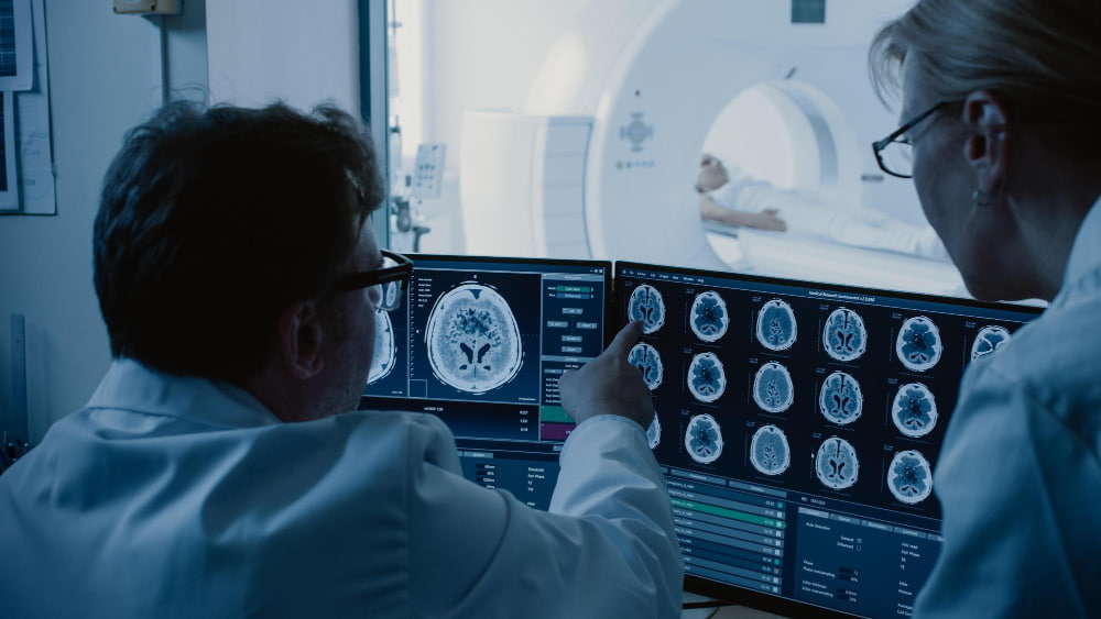

When a blood vessel supplying the brain becomes blocked, the brain's response is not uniform. Some tissue dies within minutes. Surrounding it, a larger region of brain remains electrically silent but biologically intact — still viable, still salvageable, but only if blood flow is restored in time. Identifying that salvageable tissue, and doing so fast enough to act on it, is the central challenge of acute ischemic stroke management.
Cerebral perfusion imaging exists to meet that challenge. This guide explains how cerebral perfusion imaging works, what it measures, and why it has become indispensable to modern stroke care.
What Is Cerebral Perfusion Imaging?
Cerebral perfusion imaging — also referred to as a brain perfusion scan, or brain blood flow scan — is a category of neuroimaging techniques that measure blood flow within the brain at the tissue level. Unlike standard CT or MRI, which primarily capture the structure of brain tissue, perfusion imaging reveals the physiological state of that tissue: how much blood is reaching it, how quickly, and in what volume.
In the context of acute ischemic stroke, this distinction is critical. Structural imaging can show that an infarct has occurred, but it cannot reliably show how much brain remains at risk in the surrounding territory. Perfusion imaging can. By mapping the hemodynamic state of the brain at the time of scanning, it allows clinicians to see beyond what has already been lost and identify what can still be saved.
The two main techniques used in clinical practice are CT perfusion — known clinically as CTP or perfusion CT — and MRI perfusion, also referred to as perfusion-weighted imaging (PWI). Each has distinct characteristics that make it more or less suited to specific clinical scenarios.
Cerebral Blood Flow and Its Regulation
To understand what cerebral perfusion imaging measures, it helps to understand how blood flow to the brain is normally regulated.
The brain is extraordinarily sensitive to interruptions in blood supply. It accounts for roughly two percent of body weight but consumes approximately twenty percent of the body's oxygen at rest. To protect this supply, the brain maintains a process known as cerebrovascular autoregulation — the ability to keep cerebral blood flow relatively constant across a wide range of systemic blood pressures by adjusting the diameter of small resistance vessels.
Cerebral perfusion pressure, the driving force for blood flow through the brain, is determined by the difference between mean arterial pressure and intracranial pressure. Under normal conditions, as mean arterial pressure rises or falls within a physiological range, cerebrovascular autoregulation compensates to maintain stable perfusion. This protective mechanism begins to fail when:
- Mean arterial pressure falls below a critical threshold, depriving the brain of adequate driving pressure
- Intracranial pressure rises substantially — as can occur in severe stroke, hemorrhage, or cerebral edema — compressing the cerebral vasculature and reducing effective perfusion pressure
- Autoregulatory capacity is exhausted in the setting of severe or prolonged cerebral hypoperfusion, a state of chronically or acutely inadequate blood flow to the brain resulting in a dangerous shortfall of oxygen and glucose
In acute ischemic stroke, autoregulation in the ischemic territory breaks down entirely. Cerebral blood flow in the affected region becomes passively dependent on systemic blood pressure rather than locally regulated, which is one reason blood pressure management in acute stroke requires careful, evidence-based handling rather than reflexive correction.
The Ischemic Core and Penumbra
The most important concept in cerebral perfusion imaging for stroke is the distinction between the ischemic core and the ischemic penumbra.
When a cerebral artery is occluded, blood flow drops sharply in the territory it supplies. Two distinct zones develop:
- Ischemic core — the region closest to the occlusion, where collateral circulation is absent or inadequate and blood flow falls below the threshold for cellular survival within minutes. This tissue is already irreversibly damaged at the time of imaging and cannot be salvaged regardless of how quickly reperfusion is achieved.
- Ischemic penumbra — the surrounding zone where blood flow is reduced but not absent, sustained by collateral vessels that partially compensate for the occlusion. Neurons here are functionally impaired and have stopped firing, but remain metabolically viable. This is the tissue that reperfusion therapy is designed to save.
Without intervention, the penumbra progressively recruits into the core as collateral circulation fails and cellular energy reserves are exhausted. With timely reperfusion, a substantial proportion of penumbral tissue can be rescued.
Cerebral hypoperfusion across both zones drives this process. The degree and duration of hypoperfusion determine how quickly penumbral tissue is lost, which is why time remains critical even in an era of imaging-guided patient selection.

CT Perfusion in Ischemic Stroke
CT perfusion — also known as perfusion CT or CTP — is the standard cerebral perfusion imaging technique in the acute stroke setting. It is fast, widely available, and can be performed on the same scanner immediately following non-contrast CT and CT angiography, making it a natural extension of the standard acute stroke imaging protocol.
How CT Perfusion Works
CTP is performed by injecting iodinated contrast material intravenously and acquiring a rapid series of sequential CT images over the same brain region as the contrast passes through the cerebral vasculature. The resulting time-attenuation curves are processed to generate quantitative maps of the following parameters:
- Cerebral blood flow (CBF) — the volume of blood delivered to a unit of brain tissue per unit of time, measured in milliliters per 100 grams of tissue per minute
- Cerebral blood volume (CBV) — the total volume of blood present within a unit of brain tissue at any given moment, measured in milliliters per 100 grams of tissue
- Mean transit time (MTT) — the average time taken for blood to traverse the capillary bed, which increases as perfusion pressure falls
- Time to maximum (Tmax) — the time from contrast bolus arrival to peak tissue enhancement, widely used in automated perfusion software as a marker of delayed perfusion
Identifying Core and Penumbra on CT Perfusion
CTP maps these parameters across the entire imaged brain volume, allowing the ischemic core and penumbra to be delineated:
- The ischemic core is identified by severely reduced cerebral blood volume, reflecting collapse of the local capillary bed in irreversibly damaged tissue
- The ischemic penumbra appears as a larger surrounding region of reduced cerebral blood flow and prolonged mean transit time, where the vasculature remains at least partially patent
- The perfusion mismatch is the volume difference between the full perfusion lesion and the smaller ischemic core — representing the tissue that remains salvageable
Automated software platforms such as RAPID process CTP data within minutes and generate standardized mismatch ratios and absolute volumes that feed directly into clinical decision-making at the bedside.
CT Perfusion and Treatment Selection
CTP has become central to patient selection for mechanical thrombectomy and intravenous thrombolysis, particularly in two scenarios where its contribution is greatest:
- Extended time window — between four and a half and twenty-four hours after symptom onset, clinical trials including DAWN and DEFUSE 3 demonstrated that patients with a significant perfusion mismatch could benefit substantially from mechanical thrombectomy despite the elapsed time. These trials were built on perfusion CT data and have directly changed international stroke guidelines.
- Unknown onset time — in patients who wake with stroke symptoms or cannot report when they began, the time since occlusion cannot be established from history alone. CTP provides a tissue-based assessment of salvageability that substitutes for the unavailable clock. Patients with a favorable mismatch profile are treated as if within the therapeutic window.
MRI Perfusion in Stroke
MRI-based perfusion imaging, also known as perfusion-weighted imaging or PWI, provides complementary information to CT perfusion with distinct advantages in certain clinical situations.
The most widely used MRI perfusion technique is dynamic susceptibility contrast (DSC) MRI, which tracks the passage of a gadolinium-based contrast agent through the cerebral microvasculature, generating maps of cerebral blood flow, cerebral blood volume, and mean transit time analogous to those produced by perfusion CT.
MRI perfusion is typically acquired alongside diffusion-weighted imaging (DWI), which identifies the ischemic core with high sensitivity by detecting restricted water diffusion in cytotoxically swollen tissue. The mismatch between the DWI lesion and the larger DSC perfusion deficit is the DWI-perfusion mismatch — the MRI equivalent of the CT perfusion mismatch concept.
Compared with CT perfusion, MRI perfusion offers several advantages:
- No ionizing radiation
- Simultaneous acquisition of the most sensitive available core marker (DWI) alongside the perfusion data
- Superior soft tissue contrast for characterizing the ischemic territory
- Availability of arterial spin labeling (ASL), a non-contrast technique that uses magnetically labeled arterial blood water as an endogenous tracer, suitable for serial imaging without gadolinium
Its limitations in the acute setting include:
- Longer acquisition times than CT perfusion
- More restricted availability in emergency imaging pathways
- Contraindications in patients with certain implanted devices or severe claustrophobia
- Greater sensitivity to patient movement artifact
In the subacute and chronic settings, ASL-based perfusion-weighted imaging is used for serial cerebrovascular assessment in conditions including carotid artery stenosis, chronic cerebrovascular disease, and cerebral small vessel disease, where repeated gadolinium administration is undesirable.
Perfusion Mismatch: The Basis for Treatment Decisions
Perfusion mismatch — the difference in volume between the irreversibly damaged ischemic core and the larger zone of hemodynamically compromised but viable penumbral tissue — is the central decision-making metric in imaging-guided stroke treatment.
A favorable mismatch profile is characterized by:
- A small ischemic core volume
- A large surrounding penumbra
- A high mismatch ratio between the two
This profile identifies patients in whom successful reperfusion is expected to rescue a clinically meaningful volume of brain tissue, reducing the likelihood of major disability. An unfavorable profile — a large core with little remaining penumbra — indicates that the opportunity for meaningful tissue salvage has largely passed, and that aggressive reperfusion carries a higher risk of hemorrhagic transformation without proportionate benefit.
Perfusion mismatch assessment has effectively shifted acute stroke management from a time-based paradigm to a tissue-based one. The question is no longer only how long ago the stroke occurred, but how much brain remains at risk and whether that tissue can still be saved.
Advanced Neuroimaging at Medical Park's Stroke Center
Medical Park's Stroke Center units are equipped with advanced neuroimaging technology and operates around the clock, with a multidisciplinary team of neurologists, interventional radiologists, and neurointensivists available at all hours to review imaging and make immediate treatment decisions. CT perfusion is integrated into Medical Park's standard acute stroke imaging protocol, performed on state-of-the-art CT scanners capable of whole-brain perfusion acquisition and processed using automated software that generates perfusion maps, core and penumbra volumes, and mismatch ratios within minutes. This rapid turnaround is a critical component of Medical Park's door-to-treatment workflow, supporting fast eligibility decisions for mechanical thrombectomy and intravenous thrombolysis.
For patients presenting in the extended time window or with an unclear symptom onset, perfusion CT findings directly determine treatment eligibility in line with international guidelines. MRI perfusion, including DWI-perfusion mismatch assessment and ASL-based brain perfusion scanning, is available for patients in whom MRI is clinically preferred or for subacute and chronic cerebrovascular evaluation. Medical Park's interventional neuroradiology team performs mechanical thrombectomy procedures in a dedicated neuroangiosuite, with direct integration between the imaging pathway and the intervention suite to minimize time from perfusion scan to reperfusion. Post-procedure neuroimaging, including follow-up CT and MRI perfusion studies, is used to assess reperfusion success and guide intensive care management.
International patients arriving at Medical Park with an acute stroke are received through a dedicated emergency pathway with immediate access to the full acute neuroimaging protocol. For patients referred from other centers for evaluation following a stroke, cerebral perfusion imaging is available as part of a comprehensive neuroradiological assessment coordinated across neurology, interventional neuroradiology, and neurosurgery.





 Makaleye Dön
 E-Result
 WhatsApp