


 TR
 EN
 RU
 AR
 FR


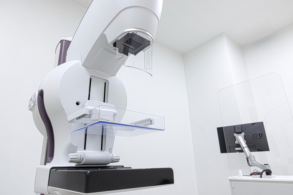

Digital breast tomosynthesis (DBT), commonly known as a 3D mammogram, is an advanced breast imaging technique used to screen for breast cancer and investigate suspicious breast findings. Unlike a standard mammogram, which produces flat, two-dimensional (2D) images, DBT captures multiple X-ray images from different angles and reconstructs them into thin sections of breast tissue. This helps radiologists examine overlapping tissue structures more clearly and may make certain abnormalities easier to detect.
A 3D mammogram is not a separate test from digital breast tomosynthesis; the terms describe the same imaging approach. Although it can improve cancer detection in some screening settings, it does not identify every breast cancer or eliminate the need for other tests when an abnormality requires further investigation.
Understanding how DBT works, its advantages and limitations, and how it compares with a standard mammogram and breast ultrasound can help you understand which imaging options may be appropriate for your breast health.
What Is Digital Breast Tomosynthesis (DBT)?
Digital breast tomosynthesis is an X-ray-based breast imaging technique that creates a series of images showing different layers of breast tissue. Commonly known as a 3D mammogram, DBT allows radiologists to examine breast tissue in sections rather than relying only on flat, two-dimensional images.
The process works as follows:
Image Acquisition: The X-ray tube moves in a small arc around the compressed breast, capturing multiple images from different angles.
Volumetric Reconstruction: A computer processes these images into thin sections through the breast tissue. This reconstruction helps reduce the effect of overlapping structures.
Image Interpretation: A radiologist examines the reconstructed sections for abnormalities such as masses, areas of architectural distortion, and suspicious calcifications.
The main difference between DBT and a standard mammogram is how the breast tissue is displayed. On a conventional 2D image, normal tissue structures may overlap and obscure an abnormality or create an appearance that requires further investigation. With DBT, the radiologist can move through the reconstructed sections to assess the same area at different depths.
Although the term “3D mammogram” is widely used, the images are reconstructed from multiple X-ray projections rather than representing a completely unrestricted three-dimensional view of the breast.
2D Mammogram and 3D Mammogram: What Is the Difference?
Both a standard mammogram and digital breast tomosynthesis use low-dose X-rays to examine breast tissue. The main distinction is that a standard mammogram produces 2D images, while DBT creates a series of reconstructed sections that help separate overlapping structures.
| Feature | Standard 2D Mammogram | Digital Breast Tomosynthesis (DBT) |
|---|---|---|
| Image format | Flat, two-dimensional images | Multiple reconstructed sections of breast tissue |
| Image acquisition | Captures images from standard angles | Captures multiple images as the X-ray tube moves through an arc |
| Tissue overlap | Overlapping structures may obscure abnormalities | Reduces the effect of overlapping tissue |
| Cancer detection | Effective for breast cancer screening and diagnostic evaluation | May detect additional cancers in some screening settings |
| Dense breasts | Dense tissue can make abnormalities harder to see | May improve the visibility of some abnormalities in dense tissue |
| Additional testing | Some findings require extra views or other imaging | May reduce screening callbacks in some populations, but further tests may still be needed |
| Radiation exposure | Uses ionizing radiation | Depends on the protocol; acquiring separate 2D and DBT images increases the total dose compared with either acquisition alone |
Neither method detects every breast cancer, and the advantages of DBT vary among patients and screening settings. The choice depends on the reason for imaging, breast density, individual risk factors, and the available equipment.
What Are the Advantages of Digital Breast Tomosynthesis?
The main advantage of DBT is its ability to reduce the visual effect of overlapping breast tissue. This can help radiologists identify and assess certain abnormalities that may be difficult to evaluate on standard 2D images.
Potential advantages include:
Improved Visibility of Breast Tissue: Examining thin sections helps radiologists distinguish some abnormalities from overlapping normal structures.
Additional Cancer Detection: DBT can identify cancers that may not be as apparent on a standard mammogram in some screening populations.
Fewer Screening Callbacks in Some Settings: By clarifying overlapping tissue, DBT may reduce the need to recall patients for additional imaging after an initial screening examination.
More Detailed Assessment of Suspicious Findings: The reconstructed images can help radiologists evaluate the location, shape, and margins of certain masses or areas of architectural distortion.
Potential Benefits for Dense Breasts: DBT may make some abnormalities easier to identify when overlapping glandular and fibrous tissue limits the visibility of conventional 2D images.
These benefits do not mean that every patient needs DBT or that it is always superior to a standard mammogram. Digital breast tomosynthesis can still produce findings that require additional tests, and some cancers may remain undetected. The decision to use it should reflect the patient's screening needs and clinical circumstances.
Dense Breasts and Digital Breast Tomosynthesis
Breast density refers to the proportion of fibrous and glandular tissue compared with fatty tissue in the breast. It is assessed on a mammogram and is not determined by breast size or how firm the breasts feel.
Dense breasts matter for two main reasons:
They Can Make Cancer Harder to Detect: Dense tissue and many breast cancers both appear white on mammograms, so an abnormality may be hidden within the surrounding tissue.
They Are Associated With a Higher Risk of Breast Cancer: Breast density is one of several factors considered when assessing a person's overall risk.
Digital breast tomosynthesis may help address the problem of overlapping tissue by allowing radiologists to examine reconstructed sections individually. This can improve the visibility of some abnormalities compared with standard 2D imaging, although dense tissue can still limit detection.
Having dense breasts does not automatically mean that additional imaging is necessary. Depending on breast density, personal and family history, and other risk factors, a healthcare provider may recommend further evaluation with ultrasound or magnetic resonance imaging (MRI).
The most appropriate screening plan depends on the individual's overall risk rather than breast density alone. DBT can be useful, but it does not replace every other breast imaging method.
Digital Breast Tomosynthesis and Ultrasound
Breast tomosynthesis and ultrasound use different technologies and answer different imaging questions. DBT uses X-rays to examine breast tissue in reconstructed sections, while ultrasound uses high-frequency sound waves to produce images of internal structures.
Digital breast tomosynthesis may be useful when:
Screening for breast cancer in an appropriate population.
Assessing an area of overlapping tissue seen on a standard mammogram.
Examining a suspicious mass or area of architectural distortion in greater detail.
Improving the visibility of some abnormalities in dense breast tissue.
Breast ultrasound may be useful when:
Investigating a palpable breast lump or a specific area of concern.
Determining whether a mass is fluid-filled, such as a simple cyst, or solid.
Examining an abnormality identified on a mammogram.
Providing supplemental imaging when clinically indicated, including in selected patients with dense breasts.
Neither examination is universally better than the other. For example, if a mammogram shows a suspicious mass, ultrasound may help characterize it. If overlapping tissue makes an area difficult to assess, DBT may provide additional information.
Ultrasound does not use ionizing radiation, but it can identify findings that require additional imaging or biopsy even when they are not cancerous. It also does not replace mammography for routine breast cancer screening in the general screening population. The choice of imaging depends on the patient's symptoms, risk factors, and the finding that needs to be investigated.
When Is Digital Breast Tomosynthesis Recommended?
DBT can be used for both cancer screening and diagnostic evaluation. The purpose of the examination influences the imaging approach and the next steps following the results.
Breast Cancer Screening
Screening is performed to look for signs of breast cancer in people who do not have symptoms. Digital breast tomosynthesis may be used for screening according to the patient's risk profile, the applicable screening recommendations, and the imaging facility's protocols.
The appropriate age to begin screening and how often to repeat it depend on factors such as age, personal and family history, and individual risk. A 3D mammogram does not remove the need for regular screening at the recommended intervals.
Investigation of an Abnormal Mammogram
If a screening mammogram identifies a finding that needs closer examination, diagnostic imaging may include DBT, additional mammogram views, or ultrasound. Tomosynthesis can help clarify whether an apparent abnormality is caused by overlapping tissue or represents a finding that needs further assessment.
Depending on the results, the radiologist may recommend:
Additional Diagnostic Images: To examine the area more closely.
Ultrasound: To characterize a mass or investigate a specific finding.
Follow-Up Imaging: After a specified interval when monitoring is appropriate.
Biopsy: If imaging cannot establish whether the abnormality is benign or malignant.
Evaluation of Breast Symptoms
A new breast lump, nipple discharge, skin changes, or a change in breast shape should be evaluated by a healthcare professional rather than left until the next routine screening appointment.
Depending on the symptom, age, and clinical findings, diagnostic imaging may include a standard mammogram, DBT, ultrasound, or another examination. Not every symptom requires a 3D mammogram, and the appropriate test should be selected according to the clinical question.
How Should You Prepare for Digital Breast Tomosynthesis?
Preparation for DBT is generally similar to preparation for a standard mammogram. A few simple steps can help the examination proceed smoothly and support accurate image interpretation.
Before your appointment:
Avoid deodorants, powders, and lotions: Do not apply these products to your breasts or underarms on the day of the examination, as they can leave marks that may interfere with image interpretation.
Wear comfortable clothing: A two-piece outfit makes it easier to remove your top and bra before the examination.
Share your medical history: Tell the healthcare team about previous breast surgery, implants, prior abnormal mammograms, or a personal history of breast cancer.
Mention breast symptoms: Explain any lump, pain, nipple discharge, or other changes so the examination can be planned appropriately.
Inform the technologist if you may be pregnant: This allows the healthcare team to assess the most appropriate imaging approach.
Bring previous breast imaging if available: Earlier mammograms and reports can help the radiologist compare findings and identify changes over time.
If you are concerned about discomfort, let the technologist know. Breast compression is necessary to obtain clear images, but correct positioning can help make the examination more comfortable.
Is Digital Breast Tomosynthesis Safe?
Digital breast tomosynthesis is generally considered safe when performed for an appropriate medical indication. Like a standard mammogram, it uses ionizing radiation, and the equipment and imaging protocol are designed to keep exposure as low as reasonably achievable while producing images suitable for evaluation.
The total radiation dose depends on the equipment and how the examination is performed. If a separate standard 2D mammogram is acquired alongside DBT, the combined dose is higher than that of a standard 2D examination alone. Some systems generate reconstructed 2D images from the DBT data, avoiding the need for a separate 2D acquisition and reducing the additional exposure.
Other limitations include:
Temporary Discomfort: Breast compression may cause brief discomfort or tenderness.
False-Positive Findings: An abnormal-looking area may turn out to be benign but still require additional imaging or a biopsy.
Missed Abnormalities: DBT cannot detect every breast cancer, particularly when a finding is difficult to visualize.
Additional Testing: Some results require ultrasound, follow-up imaging, or tissue sampling to establish a diagnosis.
If you are pregnant or think you may be pregnant, inform your healthcare provider before the examination. The benefits, risks, and need for imaging can then be assessed in light of your clinical circumstances.
Understanding Your Digital Breast Tomosynthesis Results
A radiologist reviews the images and prepares a report describing the breast tissue and any findings that require attention. The report may mention breast density, masses, calcifications, asymmetries, or architectural distortion. These terms describe what appears on the images; they do not necessarily indicate cancer.
The report may use the Breast Imaging Reporting and Data System (BI-RADS), a standardized system for classifying breast imaging findings and recommending follow-up. Depending on the result, the next step may be routine screening, additional diagnostic imaging, short-interval follow-up, or a biopsy.
A recommendation for further testing does not automatically mean that cancer is present. Additional imaging can help clarify an uncertain finding, while a biopsy may be needed to determine whether suspicious tissue is benign or malignant.
Comparing the current images with previous mammograms is also important. Changes over time can help the radiologist assess whether a finding is new, has remained stable, or needs further investigation.
Breast Imaging in Turkey at Medical Park
At Medical Park Hospitals in Turkey, digital breast tomosynthesis can help radiologists examine breast tissue in greater detail when screening for breast cancer or investigating an abnormal mammogram. By reconstructing multiple X-ray images into thin sections, this technique helps reduce the effect of overlapping tissue, which can otherwise make some abnormalities harder to see. The findings help determine whether an area needs closer examination through additional imaging or a biopsy.
Digital breast tomosynthesis is an important tool for breast cancer screening and diagnostic evaluation, but it cannot confirm or rule out cancer on its own. If imaging reveals a suspicious area, additional tests, including breast ultrasound or a biopsy, may be needed to determine whether the finding is benign or cancerous.
If breast cancer is diagnosed, imaging and biopsy results help establish the tumor’s characteristics and stage, guiding treatment decisions. Medical Park’s Radiology and Medical Oncology specialists collaborate with other relevant specialties to develop an individualized treatment plan based on the confirmed diagnosis, cancer stage, and the patient’s clinical needs.
For international patients seeking breast cancer screening in Turkey, Medical Park’s International Patient Services Center can help arrange appointments and explain the available options for breast imaging at the selected hospital. Patients who need further investigation after an abnormal screening result can also receive assistance arranging additional tests and specialist consultations.


 Makaleye Dön
 E-Result
 WhatsApp