


 TR
 EN
 RU
 AR
 FR


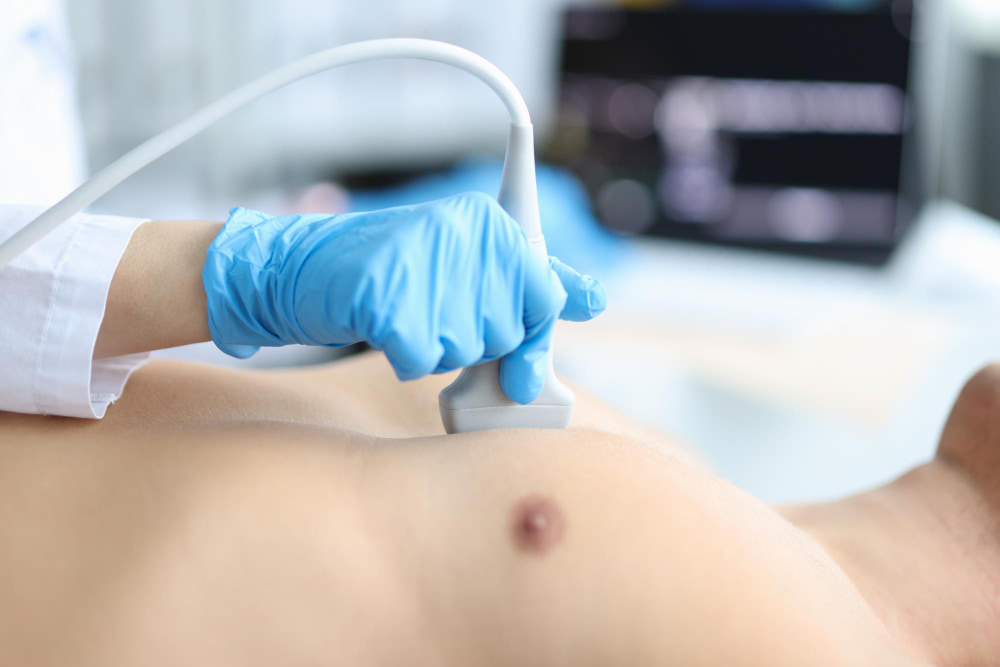


An echocardiogram is one of the most widely used and informative tests in cardiology, offering a detailed, real-time look at the heart's structure and function without a single incision or dose of radiation. For patients being evaluated for anything from a heart murmur to unexplained breathlessness, it is often the test that provides the clearest answers.
Because the heart is constantly moving, a static image can only tell part of the story. Echocardiogram, or echocardiography, captures the heart in motion, showing how its chambers fill and contract, how its valves open and close, and how blood flows through it with every beat. This guide explains what an echocardiogram involves, how it differs from an EKG, the various types of echo tests available, what the results reveal, and what patients can expect from the procedure at Medical Park.
What Is an Echocardiogram (Echocardiography)?
An echocardiogram — also called heart ultrasonography or a heart ultrasound — uses high-frequency sound waves to produce moving images of the heart's structure and function. A small device called a transducer emits sound waves that bounce off the heart's tissues and blood, and a computer converts the returning echoes into a live image displayed on a monitor.
Unlike an X-ray or CT scan, an echocardiogram test involves no ionizing radiation, making it safe for repeated use and suitable for patients of all ages, including infants and pregnant women. It is painless, non-invasive in its most common form, and typically completed within an outpatient visit. Cardiologists rely on echocardiography to assess the size and shape of the heart, the thickness and movement of the heart muscle, the condition of all four heart valves, and the direction and speed of blood flow through the heart's chambers.
Echocardiogram vs. EKG: What's the Difference?
One of the most common points of confusion for patients preparing for cardiac testing is the difference between an echocardiogram and an EKG (electrocardiogram). While both are non-invasive tests used to evaluate heart health, they measure entirely different things and are often performed together as complementary investigations.
An EKG records the heart's electrical activity — the signal that triggers each heartbeat — using electrodes placed on the skin. It is fast, typically taking only a few minutes, and is highly effective at detecting arrhythmias, conduction abnormalities, and evidence of a prior heart attack. However, an EKG cannot show the physical structure of the heart.
An echocardiogram, by contrast, uses ultrasound to visualize the heart's anatomy directly. It shows chamber size, wall thickness, valve function, and pumping efficiency — information an EKG simply cannot provide. In the echocardiogram and EKG comparison, neither test replaces the other; a cardiologist frequently orders both together, using the EKG to assess the heart's rhythm and the echocardiogram to assess its structure, to build a complete clinical picture.
When an EKG identifies an irregular rhythm such as atrial fibrillation, an echocardiogram often follows to check for structural causes or complications, and further evaluation may include stroke risk screening or treatment options such as cardiac ablation.
Types of Echocardiograms
Not every echocardiogram is performed the same way. The specific type of echocardiogram recommended depends on what a cardiologist needs to evaluate, and several distinct approaches are used in clinical practice.
Transthoracic Echocardiogram
A transthoracic echocardiogram (TTE) is the standard and most common form of the test. The transducer is placed directly on the chest wall, and images of the heart are captured through the chest from several different angles. It requires no sedation, no needles, and no special preparation, making it the first-line echocardiogram procedure for most cardiac evaluations.
Transesophageal Echocardiogram
A transesophageal echocardiogram (TEE) provides a closer, more detailed view of the heart by passing a small, flexible probe down the esophagus, which sits directly behind the heart. Because sound waves don't have to travel through the chest wall, muscle, or lung tissue, image quality is significantly sharper — particularly for structures such as the heart valves, the left atrium, and the aorta. A TEE is typically reserved for cases requiring greater precision, such as evaluating a suspected infection of a heart valve, checking for blood clots before certain cardiac procedures, or assessing complex valve disease. Patients are given mild sedation and a numbing agent for the throat, and the test is usually completed within 20 to 45 minutes.
Stress Echocardiogram
A stress echocardiogram combines heart ultrasound imaging with physical exertion, typically walking on a treadmill or, for patients unable to exercise, a medication that simulates the heart's response to stress. Images are taken before and immediately after exertion to see how the heart performs under increased demand. This test is particularly useful for detecting coronary artery disease, since reduced blood flow to the heart muscle may only become apparent when the heart is working harder.
Fetal Echocardiogram
A fetal echocardiogram evaluates the heart of a baby still in the womb, typically performed between 18 and 24 weeks of pregnancy when there is a family history of congenital heart disease, an abnormal finding on a routine prenatal ultrasound, or certain maternal risk factors. It allows specialists to detect structural heart abnormalities before birth, giving families and medical teams time to plan for delivery and, where needed, early intervention.
Echocardiogram Technology: 2D, 3D, Doppler, and Color Flow Mapping
Modern echocardiogram uses several imaging modes, often combined within a single test, to build the most complete picture of heart function possible.
- 2D echocardiogram: The foundational imaging mode, producing cross-sectional, two-dimensional views of the heart's chambers and walls in real time. This is the basis of most standard echocardiogram tests and is used to assess chamber size, wall motion, and overall structure.
- 3D echocardiogram: A more advanced technique that reconstructs three-dimensional images of the heart, offering a more precise view of valve anatomy and chamber volume. It is especially valuable for planning valve repair or replacement procedures.
- Doppler echocardiogram: Uses the Doppler effect to measure the speed and direction of blood flow through the heart's chambers and valves, helping identify abnormal flow patterns such as a narrowed or leaking valve.
- Color flow mapping: A form of Doppler imaging that overlays flow information onto the 2D image in color, allowing a cardiologist to instantly visualize the direction of blood flow and quickly spot turbulence, leakage, or obstruction that might otherwise require more detailed analysis to detect.
What Does an Echocardiogram Show?
An echocardiogram provides a comprehensive assessment of both the structure and the function of the heart, and the findings guide a wide range of clinical decisions. Among the most important measurements is ejection fraction — the percentage of blood pumped out of the heart's main pumping chamber with each contraction. An echocardiogram for ejection fraction is one of the most frequently ordered cardiac tests, since a reduced ejection fraction is a key indicator of heart failure and helps guide treatment decisions.
Beyond ejection fraction, an echocardiogram can reveal:
- Valve function: Whether heart valves are opening and closing properly, or are narrowed (stenosis) or leaking (regurgitation).
- Chamber size and wall thickness: Enlargement or thickening of the heart muscle, which can indicate conditions such as hypertrophic cardiomyopathy or long-standing high blood pressure.
- Pumping function: How efficiently the heart contracts and relaxes with each beat.
- Blood clots or masses: Abnormal structures within the heart's chambers, a finding of particular concern in patients with atrial fibrillation, since clots that form in the heart are a leading cause of ischemic stroke.
- Fluid around the heart: A buildup of fluid in the sac surrounding the heart, known as a pericardial effusion.
- Congenital abnormalities: Structural heart defects present from birth, in both children and adults.
The Echocardiogram Procedure: What to Expect
For a standard transthoracic echocardiogram, the procedure is straightforward and requires no special preparation. Patients change into a gown and lie on an exam table, usually on their left side to bring the heart closer to the chest wall. A small amount of gel is applied to the skin to improve sound wave transmission, and the sonographer moves the transducer across several positions on the chest to capture images from multiple angles.
How long does an echocardiogram take is one of the most common questions patients ask beforehand. A standard transthoracic echocardiogram typically takes between 30 and 60 minutes, depending on the complexity of the images required and whether Doppler or color flow mapping is included. A transesophageal echocardiogram usually takes 20 to 45 minutes, plus recovery time after sedation. A stress echocardiogram, which includes both resting and post-exercise imaging, generally takes 45 to 60 minutes in total. In all cases, the test itself is painless, and most patients are able to resume normal activities immediately afterward, with the exception of TEE patients, who are advised not to drive due to sedation.
Advanced Cardiac Screning and Diagostics at Medical Park
Turkey has become a leading destination for patients seeking fast, high-quality cardiac screening and diagnostics, and an echocardiogram in Turkey at Medical Park reflects that reputation. Rather than facing the waiting lists common across much of Western Europe, patients can typically arrange and complete a transthoracic, transesophageal, stress, or fetal echocardiogram within a single visit, often as part of a broader cardiovascular screening program.
Medical Park's Cardiology departments are equipped with advanced, internationally calibrated ultrasound systems capable of full 2D, 3D, Doppler, and color flow mapping imaging, and every study is interpreted by subspecialist cardiologists trained to the same standards applied across Europe and the United States.
This depth of imaging capability means an echocardiogram performed at Medical Park is rarely an isolated test — it is typically integrated with the hospital's wider suite of cardiac diagnostics, including ECG, coronary CT angiography, and cardiac MRI, allowing physicians to move seamlessly from a structural finding on ultrasound to any further imaging required for a complete diagnosis.
Patients undergoing a full body check-up can also have an echocardiogram included as part of a wider preventive health care assessment, giving physicians a complete picture of cardiovascular health alongside other routine screenings.
For international patients, Medical Park's International Patient Center manages the entire visit from first enquiry onward, including appointment scheduling, multilingual support, and travel and accommodation coordination. Patients receive a full written report in English, along with digital copies of their imaging studies, so results can be shared with a physician back home.
Whether the goal is a routine echocardiogram to investigate a new symptom or a more advanced cardiac imaging evaluation, Medical Park is positioned to deliver accurate, timely results without the delays often associated with cardiac testing elsewhere.


 Makaleye Dön
 E-Result
 WhatsApp