


 TR
 EN
 RU
 AR
 FR


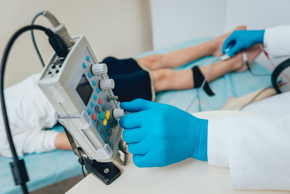

Electromyography (EMG) is a diagnostic test that records electrical activity in muscles to help identify the cause of weakness, numbness, tingling, cramps, or pain. It is usually performed alongside a nerve conduction study (NCS), which measures how well electrical signals travel through nerves.
Together, these tests help doctors determine whether symptoms may be related to a nerve problem, a muscle disorder, or the connection between nerves and muscles. Although the examination can cause temporary discomfort, it is generally safe and can provide important information for diagnosis and treatment planning.
This guide explains what an EMG test is, what conditions it can help diagnose, how to prepare for the procedure, what to expect during the examination, and how to understand the results.
What Is Electromyography (EMG)?
Electromyography, commonly called EMG, measures the electrical activity produced by muscles at rest and during contraction. Muscles depend on signals from nerves to move, and problems affecting these signals can lead to weakness, changes in sensation, or difficulty controlling movement. An EMG test records the muscles' electrical responses to help identify patterns associated with nerve or muscle dysfunction.
During needle EMG, a specialist inserts a thin electrode into selected muscles. An electrode is a device that detects electrical activity, which appears as waveforms on a screen and may also be heard as sounds through the equipment. The specialist examines how the muscle behaves at rest and when you contract it, looking for changes that may help explain your symptoms.
The term EMG test often refers to an appointment that includes both needle EMG and a nerve conduction study. These are separate but complementary tests, and each provides different information about how the nerves and muscles are working.
EMG vs. Nerve Conduction Study
A nerve conduction study (NCS) measures how quickly and effectively electrical signals travel along peripheral nerves. Small electrodes placed on the skin deliver brief electrical pulses and record the resulting responses. Needle EMG, by comparison, records electrical activity within selected muscles.
| Feature | Needle EMG | Nerve conduction study (NCS) |
|---|---|---|
| What it measures | Electrical activity within muscles | Electrical signals traveling along nerves |
| How it is performed | A thin needle electrode is inserted into selected muscles | Electrodes on the skin deliver brief electrical pulses and record responses |
| What it helps assess | Muscle disorders and changes in muscles caused by nerve problems | Nerve compression, peripheral neuropathy, and other forms of nerve dysfunction |
| What it feels like | A pinprick or aching sensation | Brief tingling or shock-like sensations |
The tests often work best together. NCS helps assess nerve function, while needle EMG shows how muscles supplied by those nerves are responding. The specialist decides which nerves and muscles to examine based on your symptoms and physical examination.
Other Types of EMG
Needle EMG is the main diagnostic form used to investigate suspected nerve and muscle disorders. Surface EMG uses electrodes attached to the skin and is used in research, movement analysis, and biofeedback therapy, including some pelvic floor rehabilitation programs. It is not a substitute for diagnostic needle EMG.
Single-fiber EMG is a specialized test that assesses communication between nerves and individual muscle fibers. It may be used when a neuromuscular junction disorder, such as myasthenia gravis, is suspected and further testing is needed.
Why Is an EMG Test Done? What Can It Diagnose?
A doctor may recommend an EMG test when symptoms suggest that nerves or muscles are not functioning as expected. Common reasons include persistent numbness, tingling, muscle weakness, unexplained cramps, and pain that may originate from a nerve. The test helps narrow down possible causes, but it does not diagnose every condition by itself or guarantee that the source of symptoms will be identified.
Nerve Compression and Radiculopathy
Nerve compression occurs when pressure affects a nerve and interferes with its function. Carpal tunnel syndrome is one example: the median nerve is compressed at the wrist, potentially causing numbness, tingling, pain, or weakness in the hand. Nerve conduction studies can help identify changes in the median nerve and assess their severity, while needle EMG may help determine whether the problem has affected muscles.
An EMG can also help investigate radiculopathy, a condition involving a nerve root where it leaves the spine. A herniated disk or changes associated with cervical spondylosis may irritate or compress a nerve root, causing symptoms that extend into an arm or leg. EMG findings may help identify the pattern of nerve involvement, while an MRI may be needed to show the structural cause.
Peripheral Neuropathy and Nerve Injury
Peripheral neuropathy refers to damage or dysfunction in nerves outside the brain and spinal cord. It can cause tingling, burning sensations, numbness, or muscle weakness, often affecting the hands and feet. Diabetes is a common cause for neuropathy (diabetic neuropathy), but vitamin deficiencies, certain medications, alcohol use, inherited disorders, and other medical conditions can also contribute.
Nerve conduction studies and EMG can help determine which nerves are affected and whether the findings suggest a localized problem or a more widespread neuropathy. The tests may also help assess nerve function after an injury. Timing matters, however: some abnormalities in muscle electrical activity take around two to three weeks to become detectable after an acute nerve injury. Earlier testing may still be useful, depending on the injury and the clinical question.
Muscle Disorders
An EMG test may help investigate myopathy, a general term for a disorder affecting muscle tissue. Possible causes include inflammatory muscle diseases such as myositis, inherited conditions such as muscular dystrophy, and certain medication-related muscle problems. Symptoms may include difficulty climbing stairs, lifting the arms, or rising from a chair.
The electrical patterns recorded during needle EMG may suggest whether weakness is related to a muscle disorder rather than a problem with the nerve supplying the muscle. However, EMG cannot identify every muscle disease or establish its exact cause on its own. Blood tests, genetic testing, imaging, or a muscle biopsy may be needed.
Motor Neuron and Neuromuscular Junction Disorders
EMG can contribute to the evaluation of disorders affecting motor neurons, the nerve cells that control voluntary muscle movement. These include amyotrophic lateral sclerosis (ALS), a progressive disease affecting motor neurons. An EMG may show changes consistent with motor neuron damage, but an abnormal result does not automatically mean that a person has ALS. Diagnosis depends on the pattern of findings, symptoms, examination results, and assessment of other possible causes.
The test may also help investigate disorders of the neuromuscular junction, the point where a nerve communicates with a muscle. Myasthenia gravis is one example. Specialized examinations, including single-fiber EMG, may be used when routine testing does not provide enough information.
What an EMG Cannot Show
An EMG test does not directly examine the brain or spinal cord and cannot show the structure of a compressed nerve or herniated disk. MRI and other imaging tests provide different information by showing anatomical changes that may explain symptoms.
A normal EMG also does not rule out every nerve disorder. Small fiber neuropathy, which affects nerve fibers involved in pain, temperature, and some automatic body functions, may not be detected by routine nerve conduction studies and needle EMG. Findings can also be normal if the affected muscles were not tested or the examination was performed too early. A normal result should therefore be interpreted alongside your symptoms and physical examination.
How to Prepare for an EMG Test
Most people do not need extensive preparation for an EMG test. You can usually eat, drink, and take your regular medications as usual unless your healthcare team gives you different instructions. Before the appointment, make sure the examiner knows about your medical conditions, medications, and any implanted electrical devices.
What to Tell Your Doctor
Blood thinners or bleeding disorders: These can increase the risk of bleeding or bruising after needle insertion. Do not stop prescribed medication unless the clinician managing your care specifically instructs you to do so.
Pacemakers or other implanted electrical devices (CIEDs): Tell the examiner before the nerve conduction study so the appropriate precautions can be considered.
Previous lymph node removal: If you have had lymph nodes removed from an armpit, ask the examiner whether needle testing in that arm should be avoided.
Recent nerve injuries: Tell the doctor when the injury occurred and how it happened, as this may affect the timing and interpretation of the test.
Planned creatine kinase (CK) blood tests: Needle EMG can temporarily raise CK, an enzyme measured when muscle damage is suspected. Ask whether the blood sample should be taken before the examination.
On the Day of the Test
Shower beforehand and avoid applying lotions, creams, oils, or powders to the areas being examined, as these products can interfere with skin electrodes. Wear loose clothing that allows easy access to your arms or legs, and bring a list of your medications.
Cold hands or feet can affect some nerve conduction measurements, so the examiner may warm the area before testing. If you are concerned about discomfort, let the team know before the examination begins. Children can also undergo EMG when indicated, with additional explanation and support appropriate to their age and needs.
What Happens During an EMG Test?
An EMG test is generally performed in an outpatient setting, and you can usually go home afterward. The examiner selects the nerves and muscles to test based on your symptoms and physical examination. In many cases, the nerve conduction study comes first, followed by needle EMG, although the order can vary.
The Nerve Conduction Study
The examiner places small electrodes on your skin and delivers brief electrical pulses through stimulating electrodes positioned over selected nerves. The equipment records the resulting responses to assess how quickly and effectively signals travel along the nerves.
You may feel short tingling, tapping, or shock-like sensations. The pulses last only a brief moment, and the examiner may test several nerves at different points to investigate your symptoms.
The Needle EMG
For needle EMG, the examiner inserts a thin, sterile needle electrode into selected muscles. You will usually be asked to relax the muscle first and then contract it gently, for example by moving your hand or lifting your foot. The electrode records electrical activity during rest and movement.
You may hear clicking or crackling sounds from the equipment as the muscle activity is recorded. These sounds are part of the measurement and do not indicate a problem. The number of muscles tested depends on the symptoms being investigated and the findings during the examination.
How Long Does an EMG Test Take, and Who Performs It?
A combined EMG and nerve conduction study often takes around 30 to 60 minutes, although a more extensive examination may take longer. The duration depends on how many nerves and muscles need to be assessed.
The examination is performed by a neurologist or another physician trained in electrodiagnostic testing, sometimes with assistance from trained clinical staff. The physician interprets the results alongside your symptoms, medical history, and physical examination.
Is an EMG Test Painful?
An EMG test can be uncomfortable, but most people tolerate it without difficulty. During the nerve conduction study, the electrical pulses feel like brief, sometimes strong tingling shocks. During needle EMG, insertion may feel like a pinprick followed by a deeper ache or brief discomfort when the muscle contracts.
The level of discomfort varies depending on the muscles tested and your individual sensitivity. Tell the examiner if the pain becomes difficult to tolerate so they can explain the next step and consider adjustments where possible.
After the test, the muscles may feel tender for a few days, and small bruises can appear at needle insertion sites. Most people can drive and return to normal activities afterward unless their clinician advises otherwise.
Risks and Side Effects of an EMG Test
EMG and nerve conduction studies are generally safe, and serious complications are uncommon. The main side effects of needle EMG are temporary soreness, minor bleeding, or bruising at the insertion sites. Infection is rare when the test is performed using appropriate sterile technique.
Needle placement in muscles near the chest wall carries a very small risk of pneumothorax, a condition in which air enters the space around a lung and causes it to collapse.
Seek urgent medical attention if you develop chest pain or shortness of breath after an EMG test. Contact your healthcare provider promptly if you experience increasing swelling, severe or worsening pain, persistent bleeding, fever, or spreading redness around a needle site. These symptoms are not typical of routine post-test soreness and need medical assessment.
Your examiner can explain any additional precautions relevant to your medical history, particularly if you take blood thinners or have a bleeding disorder or implanted electrical device.
Understanding Your EMG Results
An EMG report describes the electrical activity recorded from the tested muscles and, when performed, the findings from the nerve conduction study. The specialist looks at the pattern across the nerves and muscles examined and considers how it fits your symptoms, medical history, and physical examination. An EMG result is not a diagnosis on its own; it helps narrow down possible causes and guide decisions about further testing or treatment.
Normal and Abnormal EMG Results
A normal result means the examination did not identify significant abnormalities in the nerves and muscles tested. This can be reassuring, but it does not exclude every possible cause of symptoms, particularly conditions that routine EMG and nerve conduction studies cannot detect.
An abnormal result means the recordings show changes that may indicate nerve dysfunction, muscle disease, or a problem with communication between nerves and muscles. The report may describe where the changes were found and what pattern they suggest. An abnormal finding does not automatically mean a condition is severe or progressive; its significance depends on the type and extent of the changes and the clinical context.
Neuropathic and Myopathic Patterns
A neuropathic pattern suggests that the problem may involve a nerve supplying the muscle. Depending on the findings, the pattern may be consistent with nerve compression, a nerve-root problem, or a more widespread peripheral neuropathy.
A myopathic pattern suggests that the muscle itself may be affected. Neither pattern necessarily identifies the exact disease, so further investigations, such as blood tests, imaging, genetic testing, or occasionally a muscle biopsy, may be needed.
When the Result Is Normal
Symptoms can persist even when an EMG is normal. The test may not detect small fiber neuropathy, and it may miss abnormalities if the relevant nerves or muscles were not tested or if the examination occurred before changes became detectable. After an acute nerve injury, some abnormalities take two to three weeks to develop, so repeat testing may sometimes be considered.
If weakness, numbness, or pain continues, your doctor may recommend further assessment rather than assuming that the symptoms have no physical cause.
What Happens Next?
The next step depends on the findings and the reason the test was ordered. Your doctor may recommend an MRI to examine the spine or other structures, blood tests to investigate metabolic or inflammatory causes, or additional specialist testing. If the results support a particular diagnosis, they can help guide treatment and rehabilitation.
Some clinics discuss preliminary findings on the day of the examination, while the finalized report may take longer depending on the complexity of the test and the clinic's process. Ask your care team when to expect your results and who will explain them to you.
EMG vs. MRI and Other Tests
An EMG test measures electrical activity in nerves and muscles, while imaging and laboratory tests examine different aspects of health. Doctors may recommend more than one investigation because no single test can answer every question about the cause of weakness, pain, or numbness.
MRI: Produces detailed images of structures such as the spine, brain, and soft tissues, helping identify changes such as a herniated disk.
CT scan: Provides detailed images of internal structures, particularly bones and certain tissues.
Blood tests: Help investigate possible metabolic, inflammatory, hormonal, or muscle-related causes.
Muscle biopsy: Examines a small sample of muscle tissue when a muscle disease requires further investigation.
For example, an EMG may suggest that a nerve root is affected, while an MRI may show a herniated disk pressing on that nerve. The tests provide complementary information, and your doctor will recommend them according to your symptoms and examination findings.
Neuromuscular Testing and Neurological Care at Medical Park
At Medical Park hospitals in Turkey, neuromuscular testing helps investigate symptoms such as muscle weakness, numbness, tingling, persistent cramps, and unexplained changes in movement or muscle function. Specialists assess the patient's symptoms, medical history, and physical examination findings to determine whether electromyography (EMG) and nerve conduction studies are appropriate.
These tests evaluate electrical activity in muscles and how effectively nerves transmit signals, helping identify patterns associated with nerve compression, peripheral nerve damage, nerve root disorders, and muscle disease. Depending on the results, further investigations, including blood tests or imaging, may be recommended to clarify the diagnosis and guide treatment.
The findings of neuromuscular testing help shape the patient's care plan by showing whether symptoms are more consistent with a nerve disorder, a muscle disorder, or another underlying cause. Medical Park's Neurology specialists interpret EMG and nerve conduction study results alongside clinical findings to determine whether additional testing, medical treatment, or further specialist assessment is needed.
When a condition affects muscle strength, mobility, or daily activities, specialists in Physical Therapy and Rehabilitation can help develop a rehabilitation plan suited to the patient's diagnosis and functional needs. Depending on the condition, ongoing care may include medication, rehabilitation, or clinical monitoring.
Medical Park’s International Patient Services Center helps international patients coordinate Neurology consultations, EMG and nerve conduction study appointments where available, and any further assessments recommended by their care team. The center can also assist with treatment planning and follow-up arrangements in Turkey, helping patients navigate the next steps after their evaluation.


 Makaleye Dön
 E-Result
 WhatsApp