


 TR
 EN
 RU
 AR
 FR


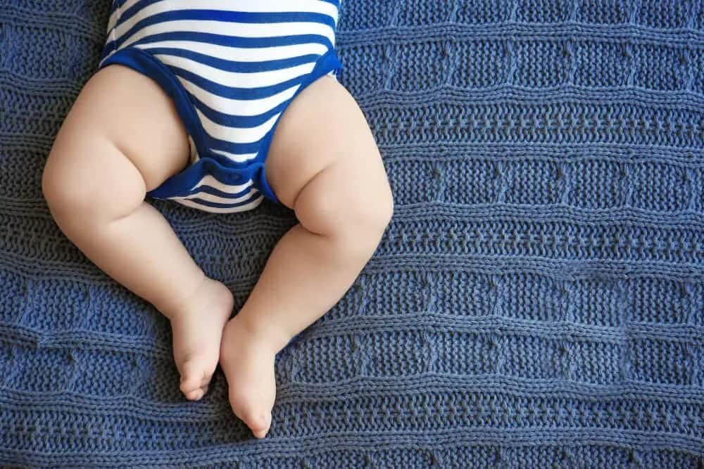


Hip dysplasia, also known as developmental dysplasia of the hip (DDH) is a structural condition in which the hip joint does not develop correctly: the ball of the femur fails to sit properly within the hip socket, leading to instability, abnormal joint loading, and progressive damage over time. It is one of the most common musculoskeletal conditions diagnosed in newborns, yet it also affects a significant number of adolescents and adults, either because it was missed in infancy or because it developed gradually over years of abnormal joint mechanics.
This guide covers everything you need to know about hip dysplasia from early signs in newborns and infants to symptoms, diagnosis, and treatment options in adults.
What Is Hip Dysplasia?
The hip is a ball-and-socket joint. In a healthy hip, the rounded head of the thigh bone (femur) sits snugly inside the cup-shaped socket of the pelvis, called the acetabulum. In hip dysplasia, this socket is too shallow, too steep, or incorrectly angled, meaning the femoral head cannot sit securely in place.
Hip dysplasia exists on a spectrum. In mild cases, the hip is simply unstable. In more severe cases, the femoral head may be partially displaced (subluxated) or fully dislocated from the socket. The condition can affect one hip or both.
Congenital Hip Dysplasia vs. Developmental Dysplasia of the Hip (DDH)
Two terms are frequently used to describe this condition, and understanding the difference matters for both parents and patients.
- Congenital hip dysplasia refers to hip dysplasia that is present at birth. It may result from the baby's position in the womb, a genetic predisposition, or hormonal factors during pregnancy that cause ligament laxity around the hip joint.
- Developmental dysplasia of the hip (DDH) is the broader, medically preferred term today. DDH encompasses cases present at birth as well as cases that develop during the first months of life as the hip continues to grow and mature. It also covers the spectrum of hip dysplasia that goes undetected and persists into adulthood.
The two terms are often used interchangeably, and DDH is now the standard clinical classification used by orthopedic specialists worldwide.
Risk Factors for Hip Dysplasia
Certain individuals are at a higher risk of developing hip dysplasia. Known risk factors include:
- Family history: A parent or sibling with hip dysplasia significantly raises the likelihood of the condition.
- Female sex: Women are four to five times more likely to be affected than men, largely due to greater sensitivity to the hormone relaxin, which loosens ligaments around the hip joint.
- Breech birth position: Babies born in the breech position (feet or bottom first) have a substantially higher risk, as the hip joint is placed under abnormal mechanical stress in the womb.
- Being first-born: The less-stretched uterus of a first-time mother may reduce fetal movement and increase hip pressure.
- Oligohydramnios: Low amniotic fluid levels restrict fetal movement and can interfere with normal hip development.
- Tight swaddling: Swaddling with the legs extended and held together has been associated with an increased risk of DDH.
- Participation in high-impact sports: In adults, repetitive loading on a mildly dysplastic hip can accelerate joint damage and bring previously silent dysplasia to clinical attention.
Signs and Symptoms of Hip Dysplasia in Babies
In many infants, DDH causes no apparent pain, which is one reason it can be easy to miss without proper clinical screening. However, there are several physical signs that parents and pediatricians should watch for.
Signs of Hip Dysplasia in Newborns and Young Infants
- Asymmetrical skin folds: Uneven creases on the thighs or buttocks, with one side showing more folds than the other.
- Limited range of motion: Difficulty spreading the legs apart, particularly during diaper changes (limited hip abduction).
- One leg appearing shorter: A visible difference in leg length when the baby is lying flat.
- A clicking or clunking sensation: A palpable or audible click in the hip joint during movement. Note that not all hip clicks indicate dysplasia.
- Unequal knee heights: When the baby lies on their back with knees bent and feet flat, one knee may appear lower than the other — a positive Galeazzi sign.
Signs of Hip Dysplasia in Older Infants and Toddlers
- A waddling or uneven gait when walking begins
- Walking on tiptoes on one side
- Limping without a clear injury
- One hip appearing higher than the other
- Leaning to one side when standing
Signs and Symptoms of Hip Dysplasia in Adults
Hip dysplasia in adults is more common than many people realize. In a significant proportion of cases, the condition was present but mild enough to go undetected in infancy and childhood, only becoming symptomatic when the joint begins to break down under the cumulative load of daily activity. In other cases, it is identified for the first time in young adults — particularly active individuals or women during or after pregnancy, when hormonal changes place additional stress on an already unstable joint.
Because adult hip dysplasia does not always cause obvious symptoms in its early stages, it is frequently misdiagnosed as a muscle strain, sports injury, or general hip pain. This delay in diagnosis often means that by the time the condition is correctly identified, significant joint damage has already occurred.
Common Symptoms of Hip Dysplasia in Adults
- Groin pain: The most characteristic symptom — a deep, aching pain in the front of the hip or groin, often worsening with activity, prolonged walking, or extended periods of sitting.
- Hip instability: A sensation of the hip clicking, catching, giving way, or feeling loose during movement.
- Pain at the end range of motion: Discomfort when rotating the hip inward or when bringing the knee toward the chest.
- Activity-related pain: Pain that begins or intensifies during running, climbing stairs, or impact sports, and may persist as an ache at rest afterward.
- Night pain: In more advanced cases, hip pain that interrupts sleep.
- Limping or altered gait: Subtle changes in the way a person walks, often developing gradually and sometimes only noticed by others.
- Early-onset hip osteoarthritis: Joint degeneration occurring in patients in their 20s, 30s, or 40s — far earlier than typical arthritis. This is one of the most serious long-term consequences of untreated hip dysplasia.
- Labral tears: Damage to the ring of fibrocartilage surrounding the hip socket, which frequently accompanies dysplasia and causes sharp, stabbing pain with specific movements.
Hip Dysplasia in Adults: Who Is Most Affected?
Hip dysplasia in adults disproportionately affects women, typically between the ages of 20 and 40. It is a leading cause of hip pain in young active women and a major driver of premature hip replacement in this age group. Athletes — particularly runners, dancers, and those involved in sports requiring repetitive hip rotation — are also at elevated risk of developing symptoms from a previously silent dysplastic hip.
Pregnancy can unmask or worsen previously undiagnosed hip dysplasia, as the hormone relaxin causes generalized ligament laxity, further destabilizing an already shallow joint. Women who experience persistent hip or groin pain during or after pregnancy should be evaluated for underlying dysplasia.
How Is Hip Dysplasia Diagnosed?
Diagnosis in Newborns and Infants
In newborns, diagnosis begins with a physical examination. Two standard clinical tests are used:
- Barlow maneuver: Checks whether a hip that is in position can be dislocated.
- Ortolani maneuver: Checks whether a dislocated hip can be reduced — that is, returned to its correct position.
Imaging is then used to confirm the diagnosis and assess severity:
- Ultrasound is the preferred imaging method in infants under 6 months of age, as the bones are still largely cartilaginous and do not appear clearly on X-ray. It allows direct visualization of the femoral head and acetabulum.
- X-ray becomes the primary imaging tool after approximately 4 to 6 months, once the bones have begun to ossify.
- MRI or CT scans may be used in complex or surgical cases for detailed planning.
Diagnosis in Adults
Diagnosing hip dysplasia in adults requires a combination of clinical assessment and imaging:
- Physical examination: The orthopedic specialist assesses range of motion, hip stability, and pain provocation. The anterior impingement test and apprehension test are commonly used to identify dysplasia-related instability and labral involvement.
- X-ray: The cornerstone of adult hip dysplasia diagnosis. Key measurements — including the lateral center-edge angle, acetabular index, and Tönnis angle — are used to quantify the degree of undercoverage of the femoral head.
- MRI: Essential for evaluating soft tissue structures, particularly the labrum, cartilage, and surrounding musculature. MRI arthrography (with contrast injection into the joint) provides the most detailed assessment of labral tears and cartilage damage.
- CT scan: Used for three-dimensional analysis of bony anatomy, especially in surgical planning for osteotomy procedures.
Because adult hip dysplasia so frequently mimics other conditions, a high index of clinical suspicion is essential. Any young or middle-aged adult with unexplained groin or hip pain — particularly one with a history of childhood hip problems or a family history of hip dysplasia — should undergo targeted imaging.
Hip Dysplasia Treatment
Treatment for hip dysplasia depends on the patient's age, the severity of the condition, and the degree of joint damage present. The overarching goal is to restore correct alignment of the femoral head within the acetabulum — and to do so before irreversible cartilage damage occurs.
Pavlik Harness for Hip Dysplasia
The Pavlik harness is the first-line treatment for infant hip dysplasia, used in babies from birth to approximately 6 months of age. It is a soft brace made of fabric straps that holds the baby's hips in a flexed and abducted position — placing the femoral head into the socket and maintaining that position while the surrounding structures develop and stabilize.
The harness is worn continuously — usually 23 hours per day — for 6 to 12 weeks, depending on the severity of the case and the baby's response. It allows free kicking movement, which actively stimulates acetabular development.
Key points about the Pavlik harness:
- Success rates exceed 85 to 95 percent when treatment is initiated within the first 2 to 3 months of life.
- Parents are fully trained in harness care, skin monitoring, and daily care routines.
- Regular ultrasound follow-up is required to confirm correct hip positioning and treatment progress.
- If adequate reduction is not achieved within 3 to 4 weeks, the treatment plan is reassessed.
Closed Reduction and Hip Spica Cast
If the Pavlik harness is unsuccessful, or if the baby is older than 6 months at diagnosis, a closed reduction procedure may be recommended. Performed under general anesthesia, the surgeon gently manipulates the hip to seat the femoral head correctly. An arthrogram confirms correct positioning, after which a hip spica cast — extending from the chest to below the knees — is applied and worn for 2 to 3 months.
Surgical Solutions to Hip Dysplasia
When non-surgical methods are insufficient, or when diagnosis occurs at an older age, surgery becomes necessary. The specific procedure depends on the patient's age, anatomy, and degree of joint damage.
Surgery in Children
- Open reduction: A surgical incision is made to directly access the hip, release obstructing soft tissue, and seat the femoral head correctly in the socket. This is followed by spica casting.
- Pelvic osteotomy: The acetabulum is reshaped or reoriented to provide better coverage of the femoral head. Several techniques exist depending on the child's age and anatomy.
- Femoral osteotomy: The angle or alignment of the femur is corrected to improve joint mechanics, often performed in combination with pelvic osteotomy.
Surgery in Adults
- Periacetabular osteotomy (PAO): The gold standard surgical treatment for hip dysplasia in young adults with preserved joint cartilage. The acetabulum is cut and repositioned to increase femoral head coverage, reducing pain, restoring stability, and significantly slowing the progression of joint degeneration. PAO is a major procedure with a structured rehabilitation period, but it offers the possibility of preserving the native hip joint for decades.
- Hip arthroscopy: In selected cases with mild dysplasia and labral tears without significant instability, arthroscopic surgery may be used to repair the labrum and address impingement. It is important to note that arthroscopy alone is not appropriate for significant dysplasia, and must always be considered carefully in this context.
- Total hip replacement (hip arthroplasty): When hip dysplasia has progressed to end-stage osteoarthritis with irreversible cartilage loss, total hip replacement becomes the appropriate treatment. In dysplastic hips, this is a technically more complex procedure than standard hip replacement, often requiring specialized implants and surgical techniques to reconstruct the abnormal anatomy.
The appropriate surgical approach is always determined on an individual basis, taking into account the patient's age, degree of dysplasia, cartilage condition, activity level, and long-term goals.
What Happens If Hip Dysplasia Is Left Untreated?
Untreated hip dysplasia does not resolve on its own. Without treatment, the abnormal mechanics of the joint lead to:
- Progressive cartilage wear and labral damage
- Early-onset hip osteoarthritis, often by the patient's 30s or 40s
- Chronic pain and functional limitation affecting work, sport, and daily life
- Gait abnormalities and compensatory problems in the knee, lower back, and pelvis
- The eventual need for total hip replacement at a young age
The earlier the condition is identified and treated, the greater the likelihood of preserving the native joint and maintaining full function over a lifetime.
Hip Dysplasia Screening: What Parents Should Know
Most hospitals perform a routine physical examination of every newborn's hips before discharge. However, a normal newborn exam does not entirely rule out DDH, as some cases develop or become apparent in the weeks and months that follow.
Pediatric guidelines recommend:
- A hip examination at every well-child visit during the first year of life
- Targeted ultrasound screening at 6 weeks of age for high-risk infants — those born in the breech position or with a family history of DDH
- Prompt referral to a pediatric orthopedic specialist if any abnormality is suspected
If you notice uneven skin folds, limited hip movement, asymmetrical leg length, or a limping gait when your child begins to walk, seek evaluation without delay.
Hip Dysplasia Care at Medical Park
At Medical Park, our Neonatology, Orthopedics and Pediatric Orthopedics specialists bring together extensive expertise in hip dysplasia across all age groups — from newborns identified through routine screening to active adults whose condition has gone undetected for years, and patients facing hip replacement due to advanced joint damage.
We offer:
- Newborn and infant hip screening and ultrasound evaluation
- Pavlik harness fitting and structured monitoring
- Closed reduction and hip spica casting
- Pediatric hip surgery including open reduction and osteotomy
- Periacetabular osteotomy (PAO) for young adults
- Hip arthroscopy for labral repair and impingement
- Complex total hip replacement for dysplastic hips
- Multidisciplinary rehabilitation and long-term follow-up
Every patient receives an individualized assessment and a treatment plan tailored to their anatomy, age, activity level, and long-term goals. Our aim is always the most effective, least invasive path to lasting hip health. If you are concerned about hip dysplasia in your newborn, child, or yourself, contact Medical Park today to schedule a consultation with our orthopedic specialists.




 Makaleye Dön
 E-Result
 WhatsApp