


 TR
 EN
 RU
 AR
 FR


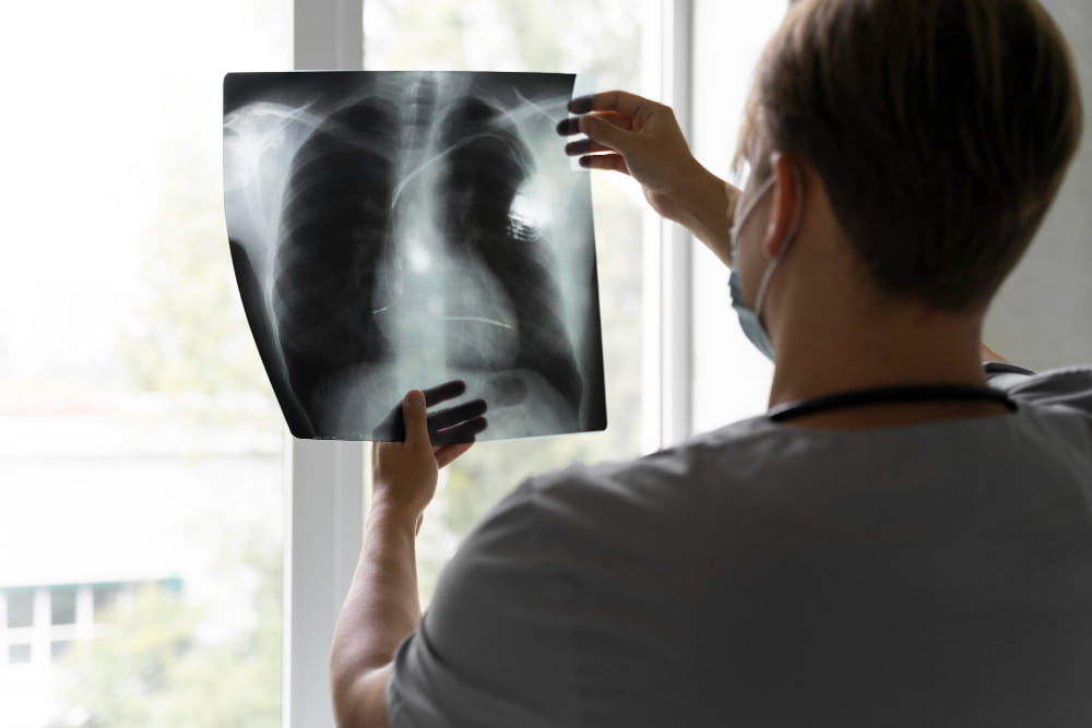

Lung Diseases: Types, Causes, Symptoms, Diagnosis, and Treatment
Lung diseases are conditions that affect the lungs and interfere with breathing or the transfer of oxygen into the bloodstream. They range from common airway conditions, such as asthma and chronic obstructive pulmonary disease (COPD), to infections, diseases that scar lung tissue, problems affecting the pulmonary blood vessels, and lung cancer.
Some develop suddenly, while others progress gradually and require long-term management. Although not every lung disease can be cured, many can be treated or controlled with appropriate medical care, monitoring, and rehabilitation. Understanding the different types, their symptoms, and how they are diagnosed can help protect lung function and reduce the risk of complications.
What Are Lung Diseases?
Lung diseases affect the structure or function of the lungs. They may interfere with airflow, the exchange of oxygen and carbon dioxide, or blood circulation through the lung tissue. Some affect a specific part of the respiratory system, while others involve several structures or develop alongside conditions affecting the heart, immune system, or other organs.
Lung diseases may be acute or chronic. Acute conditions develop over a relatively short period and may improve with treatment; pneumonia is one example. Chronic conditions, such as asthma, COPD, and pulmonary fibrosis, persist over time and may cause ongoing symptoms or gradual changes in lung function.
Lung disease does not always result from smoking or pollution. Genetic conditions, autoimmune disorders, infections, occupational exposures, and certain medications can also affect the lungs. Some people develop respiratory disease without an identifiable risk factor.
How the Lungs Work
The lungs bring oxygen into the body and remove carbon dioxide, a waste gas produced by normal cellular activity. This exchange takes place through a network of airways, air sacs, and blood vessels.
When a person breathes in, air travels through the nose or mouth and down the trachea, or windpipe. The trachea divides into two main bronchi, which branch into smaller airways called bronchioles. These passages distribute air throughout the lungs.
At the ends of the smallest airways are tiny air sacs called alveoli. Their thin walls allow oxygen to pass into nearby blood vessels, while carbon dioxide moves from the blood into the alveoli to be exhaled. Blood circulation through the lungs is therefore essential to normal gas exchange.
Lung Disease, Lung Disorders, and Pulmonary Disease
The terms lung disease, lung disorder, and pulmonary disease are generally used interchangeably. Pulmonary is the medical adjective for the lungs, while pulmonology is the specialty concerned with diagnosing and treating diseases of the lungs and respiratory system.
The term respiratory disease is broader and can include conditions affecting the nose, throat, and upper airways as well as the lungs. Some conditions also cause breathing difficulties without being primary lung diseases. For example, heart failure can cause fluid to accumulate in the lungs and lead to breathlessness, even though the underlying problem begins in the heart.
Doctors may also describe lung diseases as obstructive or restrictive. Obstructive diseases, such as asthma and COPD, make it difficult to move air out of the lungs because the airways are narrowed or obstructed. Restrictive diseases limit how much the lungs can expand, often because the lung tissue has become stiff or scarred.
Types of Lung Disease
Lung diseases can be grouped according to the structures they affect and how they interfere with breathing. The main categories include airway diseases, lung tissue diseases, lung circulation diseases, infections, lung cancer, and conditions affecting the lining around the lungs. These groups can overlap: emphysema, for example, damages the alveoli but is generally considered part of COPD.
| Category | Examples | What Happens |
|---|---|---|
| Airway diseases | Asthma, COPD, bronchiectasis, cystic fibrosis | Airways become narrowed, inflamed, obstructed, or damaged, restricting airflow |
| Lung tissue diseases | Interstitial lung disease, pulmonary fibrosis, sarcoidosis, silicosis | Lung tissue becomes inflamed, thickened, or scarred, affecting expansion and oxygen transfer |
| Lung circulation diseases | Pulmonary embolism, pulmonary hypertension, pulmonary edema | Blood flow through the lungs is blocked or restricted, or fluid accumulates in the air spaces |
| Lung infections | Pneumonia, tuberculosis, influenza, COVID-19, RSV | Microorganisms cause inflammation or infection in the airways or lung tissue |
| Lung cancer | Small cell and non-small cell lung cancer | Abnormal cells grow uncontrollably and may spread to other parts of the body |
| Pleural and chest conditions | Pleural effusion, pneumothorax, mesothelioma | The membrane around the lungs or the space between the lung and chest wall is affected |
Airway Diseases
Airway diseases affect the tubes that carry air into and out of the lungs. Inflammation, narrowing, excess mucus, or structural damage can restrict airflow and cause coughing, wheezing, chest tightness, and breathlessness.
Asthma: Asthma is a chronic condition in which the airways become inflamed and overly sensitive to certain triggers. The muscles surrounding them can tighten, while the airway lining may swell and produce extra mucus. Symptoms often vary over time and include wheezing, coughing, chest tightness, and episodes of breathlessness.
Chronic obstructive pulmonary disease (COPD): COPD causes persistent airflow obstruction and includes chronic bronchitis and emphysema. Chronic bronchitis involves long-term airway inflammation and mucus production, while emphysema damages the walls of the alveoli. Smoking is a major risk factor, although air pollution and workplace exposures can also contribute. For more information, see the dedicated COPD guide.
Bronchiectasis: This condition causes parts of the airways to become permanently widened and damaged. Mucus can collect in these areas, increasing the risk of repeated chest infections. It may develop after severe infections or alongside conditions such as cystic fibrosis.
Cystic fibrosis: This inherited condition causes thick, sticky mucus to build up in the lungs and other organs. In the lungs, mucus can obstruct airways and increase the risk of recurrent infections and progressive damage.
Alpha-1 antitrypsin deficiency: This inherited condition causes low or abnormal levels of a protein that protects lung tissue. It can lead to early-onset emphysema, including in people who have never smoked.
Bronchiolitis: This is inflammation of the small airways, most commonly affecting infants and young children. It is usually caused by a viral infection, including respiratory syncytial virus (RSV).
These conditions can share symptoms, but their causes, diagnostic findings, and treatments differ. A persistent cough or wheezing alone cannot establish which airway disease is present.
Lung Tissue Diseases
Lung tissue diseases affect the alveoli and the surrounding supporting tissue, known as the interstitium. Inflammation, thickening, or scarring can make the lungs less flexible and reduce the efficiency of oxygen transfer. Some of these conditions are described as restrictive lung diseases because they limit how fully the lungs expand.
Interstitial lung disease (ILD) is a broad group of more than 200 conditions that cause inflammation, scarring, or other changes in the tissue surrounding the air sacs. Pulmonary fibrosis refers to scarring in the lungs, and some forms of ILD cause progressive fibrosis. Idiopathic pulmonary fibrosis is a specific type in which the cause is unknown.
ILD may be associated with autoimmune diseases such as rheumatoid arthritis and systemic sclerosis. Other forms develop after exposure to silica, asbestos, certain metals, mold, or bird-related proteins. Some medications and radiation therapy can also damage lung tissue.
Symptoms include gradually worsening breathlessness, a persistent dry cough, fatigue, and reduced exercise tolerance. Diagnosis may involve lung function tests, blood tests, and high-resolution computed tomography (HRCT), a detailed type of CT scan. Blood tests may help investigate autoimmune disease. Additional tests or a lung biopsy may be considered if the diagnosis remains uncertain.
Treatment depends on the underlying condition and whether inflammation or progressive scarring is present. It may include medicines that control inflammation or immune activity, antifibrotic medicines for certain forms of progressive pulmonary fibrosis, oxygen therapy, and pulmonary rehabilitation. Avoiding an exposure responsible for the disease is also important when relevant.
Other lung tissue diseases include:
Sarcoidosis: An inflammatory condition in which small groups of immune cells, called granulomas, form in organs such as the lungs and lymph nodes. Some people have no symptoms, while others develop cough, breathlessness, chest discomfort, or fatigue.
Silicosis: An occupational disease caused by inhaling respirable crystalline silica dust. Repeated exposure can trigger inflammation and scarring, particularly in work involving stone, concrete, and sand.
Asbestosis: Lung scarring caused by prolonged inhalation of asbestos fibers. Symptoms may appear many years after exposure and include progressive breathlessness and persistent cough. Asbestos exposure is also associated with mesothelioma and lung cancer.
Hypersensitivity pneumonitis: An immune reaction in the lungs triggered by repeated inhalation of certain particles, such as mold or bird proteins. Continued exposure can cause inflammation and, in some cases, permanent scarring.
Because these conditions can have overlapping symptoms, identifying workplace, household, hobby-related, and environmental exposures is an important part of assessment.
Lung Circulation Diseases
Lung circulation diseases affect the blood vessels that carry blood through the lungs. They can reduce blood flow, interfere with oxygen delivery, or place additional strain on the heart.
Pulmonary embolism: A pulmonary embolism occurs when a blood clot blocks an artery in the lungs. The clot often travels from a deep vein in the leg or pelvis. Sudden breathlessness, chest pain, a rapid heartbeat, dizziness, or coughing up blood can occur. Suspected pulmonary embolism requires urgent medical assessment.
Pulmonary hypertension: This refers to abnormally high blood pressure in the arteries of the lungs. It can develop because of changes in the pulmonary blood vessels, heart disease, chronic lung disease, or other conditions. Over time, it may strain the right side of the heart.
Pulmonary edema: This occurs when excess fluid collects in the air spaces of the lungs. It is often associated with heart problems, particularly heart failure, and can cause severe breathlessness requiring urgent treatment.
Some lung circulation disorders overlap with cardiovascular conditions. Identifying the underlying cause is important because treatment differs between these conditions.
Lung Infections
Lung infections develop when microorganisms enter the respiratory system and cause inflammation or damage. Their severity ranges from mild illness to conditions requiring hospital treatment, particularly in infants, older adults, and people with weakened immune systems or existing lung disease.
Pneumonia: Pneumonia causes inflammation in the air sacs, which may fill with fluid or inflammatory material. It can be caused by bacteria, viruses, or fungi. Symptoms include fever, cough, chest pain, and breathlessness. The dedicated [pneumonia] guide covers its causes, diagnosis, and treatment in more detail.
Bronchitis: Bronchitis is inflammation of the bronchial tubes. Acute bronchitis is commonly caused by a virus, while chronic bronchitis involves a long-term mucus-producing cough and is often part of COPD.
Tuberculosis (TB): This bacterial infection, caused by Mycobacterium tuberculosis, most often affects the lungs. It may cause a persistent cough, fever, night sweats, fatigue, and weight loss. Treatment requires a combination of antibiotics.
Influenza: Influenza can cause fever, cough, muscle aches, and fatigue. It may lead to pneumonia or worsen existing lung and heart conditions.
COVID-19: Caused by the SARS-CoV-2 virus, COVID-19 can affect the respiratory system and sometimes cause pneumonia, reduced oxygen levels, or persistent symptoms after the initial infection.
RSV infection: Respiratory syncytial virus can cause bronchiolitis and pneumonia in infants and may lead to serious illness in older adults and people with certain underlying conditions.
Some severe respiratory infections can lead to lasting lung problems, but not everyone who develops pneumonia or a viral infection experiences chronic lung damage.
Lung Cancer
Lung cancer develops when abnormal cells in the lungs grow uncontrollably and may spread to nearby tissues or other organs. Smoking is the main risk factor, but lung cancer also occurs in people who have never smoked. Other risk factors include radon, air pollution, asbestos and other workplace carcinogens, previous radiation exposure, and family history.
The two main categories are non-small cell lung cancer, which accounts for most cases, and small cell lung cancer, which generally grows and spreads more quickly. Early lung cancer may cause no noticeable symptoms. Persistent cough, coughing up blood, chest pain, breathlessness, unexplained weight loss, and recurrent chest infections can occur, although these symptoms may have other causes.
Screening is intended for people at increased risk who do not have symptoms. In the United States, the US Preventive Services Task Force recommends annual low-dose CT screening for adults aged 50 to 80 with at least a 20 pack-year smoking history who currently smoke or quit within the past 15 years. A pack-year represents smoking an average of one pack of cigarettes per day for one year. These criteria are specific to the US recommendation and may differ in other countries. People with symptoms need diagnostic assessment rather than a screening test.
Diseases of the Pleura and Chest
The pleura is a thin, double-layered membrane that covers the lungs and lines the inside of the chest wall. Conditions affecting this membrane or the space around the lungs can cause chest pain and breathlessness.
Pleural effusion: An abnormal buildup of fluid between the pleural layers. It can occur with heart failure, infections, cancer, and other conditions. Treatment focuses on the cause and may include draining the fluid.
Pneumothorax: Also called a collapsed lung, this occurs when air enters the space between the lung and chest wall. It can develop after an injury or medical procedure or occur spontaneously. Sudden chest pain and breathlessness require urgent assessment.
Mesothelioma: A cancer that most often develops in the pleura and is strongly associated with asbestos exposure. It is distinct from lung cancer that begins in lung tissue.
What Causes Lung Disease?
Lung diseases have different causes, and more than one factor may contribute to the same condition. Common contributors include smoking, air pollution, workplace exposures, infections, genetic changes, autoimmune disorders, and certain medications.
Smoking and Tobacco
Smoking is a major risk factor for COPD and lung cancer. Tobacco smoke contains substances that irritate and damage the airways and lung tissue. Long-term exposure can contribute to chronic inflammation, excess mucus production, destruction of alveoli, and changes that increase cancer risk. Secondhand smoke can also harm the respiratory system, including during childhood when the lungs are still developing.
Quitting smoking can reduce the risk of further damage and improve health at any stage. Although it cannot reverse every change that has already occurred, it can slow the progression of smoking-related disease and lower the risk of complications. Counseling and approved smoking-cessation treatments can help people quit.
Air Pollution and Environmental Exposures
Outdoor pollution from traffic, industry, and other sources can irritate the airways and worsen existing asthma or COPD. Indoor exposures may include smoke from cooking or heating fuels, poor ventilation, mold, and dust.
Other environmental risks include:
Radon: A naturally occurring radioactive gas that can accumulate inside buildings. Long-term exposure increases lung cancer risk, including among people who have never smoked. Testing is the only reliable way to determine indoor radon levels.
Wildfire smoke: Fine particles and gases in wildfire smoke can irritate the respiratory system, particularly in people with existing lung disease.
Mold and organic particles: Repeated exposure to mold, bird proteins, and certain other particles can trigger hypersensitivity pneumonitis in susceptible people.
Vaping and e-cigarettes: E-cigarette aerosols can contain nicotine, fine particles, flavoring chemicals, and other potentially harmful substances. Their long-term respiratory effects are still being studied, but they should not be considered harmless.
Reducing exposure where possible can help protect lung health. The level of risk depends on the substance, concentration, duration of exposure, and individual susceptibility.
Workplace Exposures
Inhaling dust, fibers, fumes, or chemicals over months or years can cause occupational lung disease. Asbestos fibers can cause asbestosis and increase the risk of mesothelioma and lung cancer. Silica dust can cause silicosis, while coal dust and other mineral particles can contribute to pneumoconiosis. Certain chemical fumes can also cause occupational asthma or lung inflammation.
Ventilation, protective equipment, exposure controls, and occupational health monitoring help reduce these risks. Anyone with a history of workplace exposure should mention the type of work and approximate duration during a medical assessment, even if the exposure ended many years earlier.
Infections, Genetics, Autoimmune Disease, and Medicines
Severe respiratory infections, including pneumonia and tuberculosis, can sometimes cause lasting damage. Genetic conditions such as cystic fibrosis and alpha-1 antitrypsin deficiency can affect lung function, while autoimmune diseases such as rheumatoid arthritis and systemic sclerosis may involve the lungs as well as other organs.
Certain medications, including some chemotherapy and immune-modifying drugs, and radiation therapy involving the chest can also cause lung inflammation or damage. Doctors consider these possibilities when new respiratory symptoms develop after a medication change or treatment.
Symptoms of Lung Disease
Symptoms vary according to the condition and the part of the respiratory system affected. Some diseases cause noticeable symptoms early, while others develop gradually. Because symptoms overlap, they cannot reliably identify a specific condition without medical assessment.
Common Symptoms
Persistent cough: A cough may be dry or produce mucus. It can occur with infections, asthma, COPD, bronchiectasis, interstitial lung disease, and lung cancer.
Shortness of breath: Breathlessness may first appear during exercise and later occur during everyday activities or at rest. It can result from airway obstruction, lung tissue damage, circulation problems, or heart conditions.
Wheezing: A whistling sound during breathing can occur when the airways narrow, as in asthma or COPD.
Chest tightness or pain: Some lung conditions cause discomfort that worsens with deep breathing or coughing. Chest pain can also have cardiac or other causes.
Fatigue and reduced exercise tolerance: Breathing difficulties and reduced oxygen levels can make everyday activities more tiring.
Frequent chest infections: Repeated infections may occur with bronchiectasis, cystic fibrosis, COPD, or immune system problems.
Changes in mucus: Increased sputum, changes in its consistency, or persistent mucus production may occur with airway diseases and infections.
Clubbing of the fingers, in which the fingertips become enlarged and the nails curve more than usual, can occur in some chronic lung diseases and other medical conditions. New finger clubbing should be assessed.
Symptoms That Need Medical Assessment
Arrange a medical assessment if you experience:
A cough lasting more than two to three weeks, or one that repeatedly returns.
Breathlessness that gradually worsens or interferes with normal activities.
Unexplained weight loss, particularly alongside a persistent cough or fatigue.
Hoarseness that does not clear, especially with other respiratory symptoms.
Persistent changes in sputum, recurrent chest infections, or blood-streaked mucus.
These symptoms do not necessarily indicate a serious lung disease, but they warrant investigation. People with a history of smoking, workplace exposure, or previous lung disease should mention this when arranging an appointment.
Symptoms That Need Urgent Care
Seek emergency medical care for severe or rapidly worsening breathlessness, blue or gray lips or fingertips, significant coughing up of blood, sudden chest pain with breathlessness, confusion, fainting, or loss of consciousness. These symptoms may indicate a serious respiratory or cardiovascular problem, including pulmonary embolism or pneumothorax.
Call local emergency services if symptoms are severe rather than attempting to travel independently. Do not wait for a routine appointment.
How Are Lung Diseases Diagnosed?
Diagnosis depends on the symptoms, medical history, physical examination, and suspected condition. A doctor may begin with breathing and oxygen measurements before recommending lung function tests, imaging, laboratory investigations, or procedures to examine the airways and lung tissue.
Medical History, Examination, and Oxygen Levels
The assessment usually includes questions about symptoms, previous respiratory infections, existing conditions, medications, and family history. Doctors may also ask about smoking, secondhand smoke, workplace exposures, travel, and environmental factors such as mold or bird contact.
During the examination, a doctor listens to the chest with a stethoscope and checks breathing rate, heart rate, and other vital signs. Pulse oximetry estimates oxygen saturation using a small sensor placed on a finger. An arterial blood gas (ABG) test may be needed when more detailed information about blood oxygen levels, carbon dioxide, and blood acidity is required.
Breathing Tests
Pulmonary function tests measure how well the lungs move air and transfer gases. They help identify obstructive or restrictive patterns and monitor changes over time.
Spirometry: Measures how much air a person can breathe out and how quickly they can exhale. It is commonly used to assess asthma and COPD and may be repeated after a bronchodilator.
Lung volume testing: Measures how much air the lungs can hold and can help investigate reduced lung capacity.
Diffusion testing: Assesses how effectively oxygen passes from the air sacs into the bloodstream. It can be useful when interstitial lung disease, emphysema, or pulmonary vascular disease is suspected.
Exercise testing: A six-minute walk test measures how far a person can walk in six minutes while clinicians monitor symptoms and, when appropriate, oxygen saturation and heart rate.
Results are interpreted alongside symptoms, examination findings, and imaging. No single breathing test can diagnose every lung disease.
Imaging
Imaging tests allow doctors to examine the lungs, airways, blood vessels, and surrounding tissues.
Chest X-ray: May show pneumonia, fluid around the lungs, a collapsed lung, some tumors, and other structural changes. A normal X-ray does not exclude every lung condition.
Computed tomography (CT): Produces detailed cross-sectional images and can help identify nodules, tumors, infections, blood vessel problems, and structural changes.
High-resolution CT (HRCT): Provides detailed images of lung tissue and is particularly useful when interstitial lung disease or pulmonary fibrosis is suspected.
CT pulmonary angiography: Uses contrast material to visualize the pulmonary arteries and is commonly used when pulmonary embolism is suspected.
An imaging test may identify an abnormality, but further investigations may be needed to establish its cause.
Bronchoscopy, Biopsy, and Other Tests
Bronchoscopy involves passing a thin, flexible tube with a camera through the nose or mouth into the airways. It allows a doctor to inspect the bronchial tubes and collect mucus or tissue samples when necessary. A biopsy is the removal of a small tissue sample for examination under a microscope.
Sputum tests can identify bacteria, fungi, or other microorganisms and may be used when tuberculosis or a persistent infection is suspected. Blood tests can help investigate inflammation, autoimmune disease, and infection. Additional tests may be recommended when an inherited condition or specific immune response is suspected.
The diagnostic plan depends on the suspected condition and earlier findings. A specialist can explain the purpose of each test and whether it involves preparation, contrast material, sedation, or a recovery period.
Treatment of Lung Diseases
Treatment depends on the diagnosis, severity, rate of progression, and whether other organs are affected. Some conditions can be cured, while others require long-term management to control symptoms, prevent flare-ups, and preserve lung function. Treatment aims to address the underlying cause where possible and support breathing and everyday activity.
Medicines
Medication is selected according to the condition and the problem it causes. Common treatment groups include:
Bronchodilators: Relax the muscles around the airways and improve airflow. They are commonly delivered through inhalers for asthma and COPD.
Corticosteroids and other anti-inflammatory medicines: Reduce inflammation and may help prevent symptoms or flare-ups in selected conditions.
Antibiotics and antiviral medicines: Treat specific bacterial or viral infections when indicated. Antibiotics do not treat viral infections.
Immune-modifying medicines: May be prescribed for selected inflammatory or autoimmune lung diseases and require monitoring for side effects.
Antifibrotic medicines: Can slow the decline in lung function in some forms of progressive pulmonary fibrosis, although they do not remove existing scar tissue.
Anticoagulants: Treat pulmonary embolism and reduce the risk of further clot formation. Treatment duration depends on the cause of the clot and the person's risk of recurrence and bleeding.
Patients should use medicines as prescribed and discuss side effects or difficulties with their healthcare team rather than changing treatment independently.
Oxygen Therapy and Breathing Support
Oxygen therapy may be prescribed when testing shows persistently low blood oxygen levels. It can be delivered through a nasal cannula or mask, with the amount and duration determined by clinical assessment. Some people need oxygen during activity or sleep, while others require it for longer periods.
Oxygen is not automatically beneficial for everyone who feels breathless. Breathlessness has several possible causes, and oxygen should not be started or adjusted without medical guidance. Some people with advanced lung disease may also need noninvasive ventilation, which supports breathing through a mask. In more severe situations, invasive ventilation may be required in an intensive care unit.
Pulmonary Rehabilitation
Pulmonary rehabilitation is a supervised program for people with certain chronic respiratory conditions. It commonly combines tailored exercise, breathing techniques, education, and support with managing symptoms during daily activities.
Exercise may include walking, cycling, and strength training adapted to the person's capacity. Education can address medication use, energy conservation, nutrition, and recognizing signs of a flare-up. Rehabilitation does not replace medical treatment, but it can improve exercise tolerance and help people remain as independent as possible.
Surgery and Lung Transplantation
Surgery may be considered when a lung condition cannot be adequately managed with medication or other treatments. Thoracic surgeons perform procedures for selected lung tumors, pleural conditions, pneumothorax, and other chest diseases. The operation depends on the diagnosis, location of the problem, lung function, and overall health.
For lung cancer, surgery may be an option when the tumor can be removed and the person's health and lung function allow an operation. Other patients may need a combination of treatments, including radiation therapy or systemic medication. Decisions depend on the cancer type, stage, and individual clinical findings.
Lung transplantation may be considered for carefully selected people with severe, advanced lung disease when other treatments are no longer sufficient. It requires complex evaluation, surgery, and lifelong follow-up, including medication to prevent rejection. It is not suitable for every person with advanced lung disease.
Preventing Lung Disease and Protecting Your Lungs
Not all lung diseases can be prevented, particularly those caused by inherited conditions or factors that cannot be changed. However, reducing exposure to respiratory hazards, preventing infections, and managing existing conditions can lower the risk of some diseases and help protect lung function.
Practical steps include:
Avoid tobacco smoke: Not smoking is one of the most important ways to reduce the risk of COPD and lung cancer. Professional support and evidence-based cessation treatments can help people quit.
Reduce exposure to air pollution: Check local air quality during periods of pollution or wildfire smoke. When appropriate, reduce strenuous outdoor activity and improve indoor ventilation.
Use workplace protection: Follow safety measures when working around silica, asbestos, mineral dust, fumes, or chemicals.
Address indoor hazards: Maintain adequate ventilation, deal with persistent dampness and mold, and consider radon testing where exposure is a recognized risk.
Keep vaccinations up to date: Influenza, pneumococcal, and COVID-19 vaccination can reduce the risk of certain infections and complications. RSV vaccination is recommended for specific groups in some countries. Recommendations depend on age, health status, and local guidance.
Stay physically active: Regular activity supports general cardiovascular and respiratory fitness. People with chronic lung disease should choose activities appropriate to their symptoms.
Seek assessment for persistent symptoms: A cough that does not improve, unexplained breathlessness, or repeated chest infections should be evaluated.
Lung cancer screening may be appropriate for people who meet specific high-risk criteria. The US Preventive Services Task Force recommends annual low-dose CT for adults aged 50 to 80 with at least a 20 pack-year smoking history who currently smoke or quit within the past 15 years. Recommendations differ between countries, and low-dose CT is not a routine test for everyone. A healthcare professional can help determine whether screening is appropriate and explain its potential benefits and harms.
There is no proven supplement, drink, or commercial lung detox product that cleans or repairs damaged lungs. Protecting lung health means reducing harmful exposures, following appropriate medical advice, and managing existing conditions.
Living With a Lung Condition
A chronic lung condition may affect exercise, work, sleep, and everyday activities, but appropriate treatment and support can help many people maintain their independence. Individual needs depend on the diagnosis, severity of symptoms, and how the condition changes over time.
Following the treatment plan and attending regular reviews allow doctors to monitor symptoms, lung function, oxygen levels, and possible complications. People who use inhalers may benefit from having their technique checked. Those with a history of flare-ups should understand which changes require medical advice and which symptoms need emergency care.
Staying active within personal limits, eating a balanced diet, keeping recommended vaccinations up to date, and avoiding respiratory irritants can support general health. Breathlessness and uncertainty can also affect emotional well-being. Counseling, family support, and patient organizations may help people adjust to changes in daily life.
Pulmonology, Thoracic Surgery, and Lung Care at Medical Park
At Medical Park hospitals in Turkey, lung disease assessment and treatment may involve pulmonologists, radiologists, thoracic surgeons, medical oncologists, and physical therapy specialists, depending on the diagnosis and treatment required. Assessment may include a review of symptoms, medical and smoking history, workplace or environmental exposures, and a physical examination.
When indicated, pulmonologists use breathing tests and oxygen measurements to assess lung function. Chest X-rays, CT scans, and high-resolution CT imaging can help investigate infections, airway changes, lung nodules, and interstitial lung disease. Additional investigations, including bronchoscopy, laboratory tests, or tissue sampling, may be considered when initial findings do not establish a diagnosis.
Treatment planning depends on the condition. Pulmonologists manage medical treatment for respiratory diseases, while thoracic surgeons assess conditions that may require an operation. When lung cancer is diagnosed, oncology specialists contribute to treatment planning, and rehabilitation professionals can help address reduced exercise capacity and functional limitations.
Follow-up may involve repeat breathing tests, imaging, medication reviews, and assessment of symptoms. The specialists, diagnostic services, and treatment options available depend on the selected hospital and the patient's clinical needs.
Medical Park's International Patient Services Center helps international patients coordinate specialist consultations, diagnostic appointments, treatment planning, and follow-up arrangements in Turkey. Patients can share previous medical reports and imaging for review as part of the appointment-planning process.






 Makaleye Dön
 E-Result