


 TR
 EN
 RU
 AR
 FR


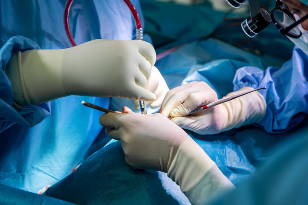

Osteotomy is a surgical procedure in which bone is deliberately cut to allow it to be repositioned, realigned, or reshaped. It is one of the most widely used techniques across surgical specialties — from orthopedic surgery and foot and ankle surgery to jaw surgery and rhinoplasty — because correcting the position of a bone can redistribute forces across a joint, address deformity, and restore function without removing the bone or replacing the joint entirely.
The word derives from the Greek osteon (bone) and tome (cutting). The procedure is defined not by a single technique but by a shared principle: that changing where and how a bone sits changes the forces acting on it, and changing those forces changes what happens to the surrounding joint, soft tissue, and the patient's symptoms.
What Is an Osteotomy?
An osteotomy involves making a precise, planned cut through bone at a specific site and in a specific direction. Once the bone is cut, it is repositioned and held in place with plates, screws, or staples while it heals in its corrected alignment. Over weeks to months, the bone unites in its new position.
Osteotomy is often confused with ostectomy, but the two procedures are distinct. An osteotomy cuts the bone and repositions it — the bone remains, just in a new alignment. An ostectomy involves removing a segment of bone entirely. The distinction matters because the two serve different clinical purposes.
Orthopedic Osteotomy
In orthopedic surgery, osteotomy is most commonly used to realign a joint affected by deformity or early arthritis, redistribute mechanical load away from a damaged area of cartilage, or preserve a joint in a younger patient for whom replacement surgery is premature. The most frequently performed orthopedic osteotomies involve the knee and hip.
High Tibial Osteotomy
High tibial osteotomy is performed in the upper tibia to shift the mechanical axis of the knee away from the damaged medial compartment and toward the healthier lateral compartment. It is most appropriate for younger, more active patients with medial compartment osteoarthritis and a varus — bow-legged — deformity who are not yet candidates for knee replacement.
The most widely used technique is the opening wedge approach, in which a wedge-shaped gap is created on the inner side of the tibia and held open with a plate, correcting the axis without removing bone. Recovery involves protected weight-bearing for approximately six to twelve weeks.
Tibial Tubercle Osteotomy
The tibial tubercle is the bony prominence on the front of the shin where the patellar tendon attaches — the connection between the quadriceps muscle and the lower leg. A tibial tubercle osteotomy detaches this prominence and repositions it to correct the tracking of the kneecap within the femoral groove. It is used to treat:
- Recurrent patellar dislocation — in which the kneecap repeatedly shifts out of its groove due to malalignment of the extensor mechanism
- Patellofemoral pain syndrome — in which abnormal kneecap tracking causes pain at the front of the knee
- Combined instability and cartilage damage — where repositioning the tubercle offloads a damaged area of the patella
The tubercle is fixed in its new position with screws.
Periacetabular Osteotomy
A periacetabular osteotomy, also known as the Ganz osteotomy, is performed around the acetabulum — the socket of the hip joint — to correct acetabular dysplasia, a common form of hip dysplasia in which the socket is too shallow or incorrectly angled to provide adequate coverage of the femoral head. This abnormal hip structure can increase pressure on the joint and place it at risk of early arthritis.
The procedure frees the acetabulum from the pelvis with a series of cuts, allowing the surgeon to rotate and tilt it into better alignment before fixing it in the new position with screws. It is generally performed in young adults before significant arthritis develops, with the goal of improving hip stability and delaying or preventing the need for hip replacement.
Femoral Osteotomy
Femoral osteotomy corrects abnormal angulation or rotation of the femur. Common indications include:
- Intertrochanteric osteotomy — used in hip dysplasia and selected cases of early avascular necrosis to unload damaged areas of the femoral head
- Distal femoral osteotomy — corrects valgus, or knock-knee, deformity when the malalignment originates in the femur rather than the tibia
Foot and Ankle Osteotomy
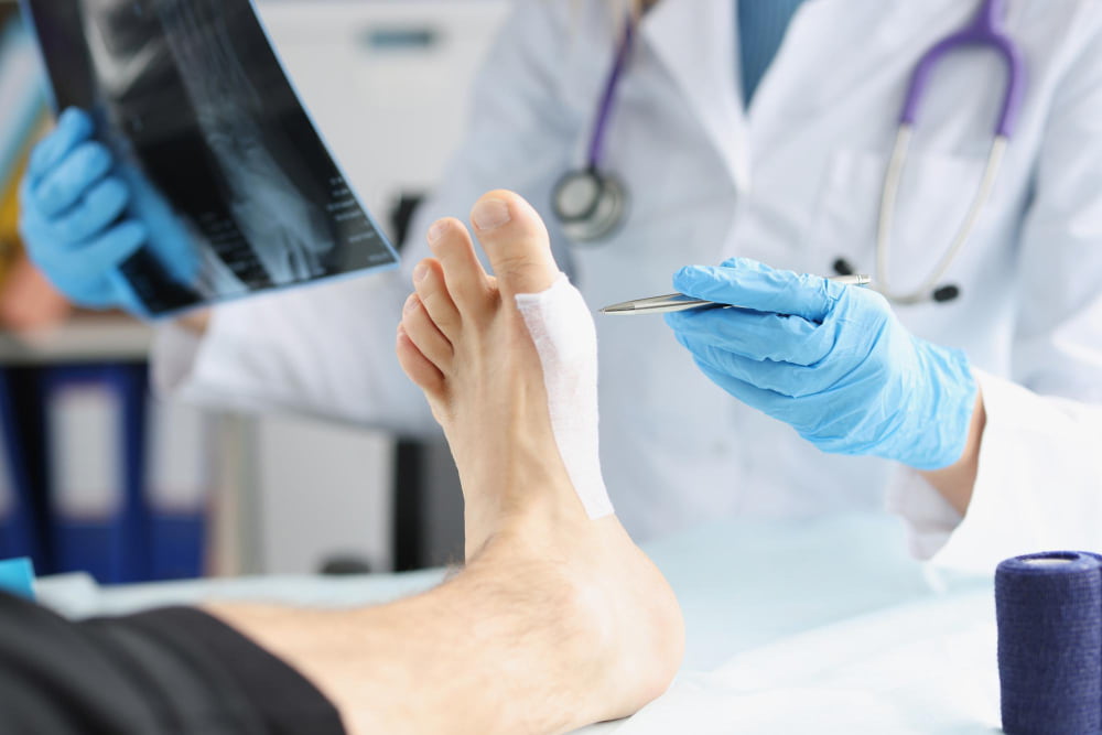
Osteotomy is used extensively in foot and ankle surgery to correct structural deformities at specific anatomical sites. Several named procedures are each associated with a particular bone, a particular deformity, and a particular technical approach.
Calcaneal Osteotomy
The calcaneus is the heel bone and the structural foundation of the hindfoot. Calcaneal osteotomy is most commonly performed as part of flatfoot reconstruction to correct hindfoot valgus — a condition in which the heel tilts outward. The medialising calcaneal osteotomy cuts obliquely through the posterior calcaneus and shifts it inward, restoring a more neutral heel position and reducing strain on the medial arch.
Hallux Valgus Osteotomies
Hallux valgus — commonly known as a bunion — is one of the most common indications for foot osteotomy. As part of bunion surgery, osteotomy may be used to realign the bones of the big toe and correct the underlying deformity. Two procedures are frequently used together:
- Chevron osteotomy — a V-shaped cut through the head of the first metatarsal that allows the metatarsal head to be shifted laterally, reducing the prominent inner border of the foot and correcting the drift of the big toe
- Akin osteotomy — a closing wedge cut through the proximal phalanx of the big toe, used alongside the chevron when the deformity has a rotational component within the toe itself
Lesser Metatarsal and Midfoot Osteotomies
Beyond the hallux, osteotomy is used to address pain and deformity in the lesser toes and midfoot:
- Weil osteotomy — a horizontal shortening cut through the head of the second, third, fourth, or fifth metatarsal, used to relieve metatarsalgia, correct plantar plate injuries, and allow lesser toe deformities to be reduced
- Cotton osteotomy — an opening wedge cut through the medial cuneiform bone in the midfoot, used in flatfoot reconstruction to plantarflex the medial column and restore arch height, typically performed alongside a calcaneal osteotomy
Jaw Osteotomy
Jaw osteotomies — formally known as orthognathic surgery — correct skeletal discrepancies between the upper and lower jaw that affect how the teeth bite together, facial balance, airway dimensions, and the ability to chew and speak comfortably. These discrepancies cannot be resolved by orthodontic treatment alone when the underlying problem is skeletal rather than dental. Jaw osteotomies are planned using three-dimensional digital imaging and surgical simulation and are carried out after a preparatory phase of orthodontic treatment.
Bilateral Sagittal Split Osteotomy
The bilateral sagittal split osteotomy is the standard operation for repositioning the lower jaw. The mandible is split along the sagittal plane on both sides, allowing the tooth-bearing segment to be advanced, set back, or rotated to correct the bite. It is used for:
- Class II malocclusion — in which the lower jaw is underdeveloped or receding relative to the upper jaw
- Class III malocclusion — in which the lower jaw is too prominent
- Facial asymmetry — where unequal jaw growth has produced imbalance between the two sides
The repositioned segments are fixed with titanium plates and screws. The jaw is functional from the day of surgery, though a soft diet is required during healing.
Premaxilla Osteotomy
The premaxilla is the anterior portion of the upper jaw carrying the four upper incisor teeth. Premaxilla osteotomy separates this segment from the rest of the maxilla to allow its independent repositioning. It is used most commonly in the surgical management of cleft lip and palate, where the premaxilla is often protruded and rotationally displaced, and in selected cases of anterior skeletal discrepancy where the front teeth and their supporting bone need to be moved as a single unit.
Rhinoplasty Osteotomy
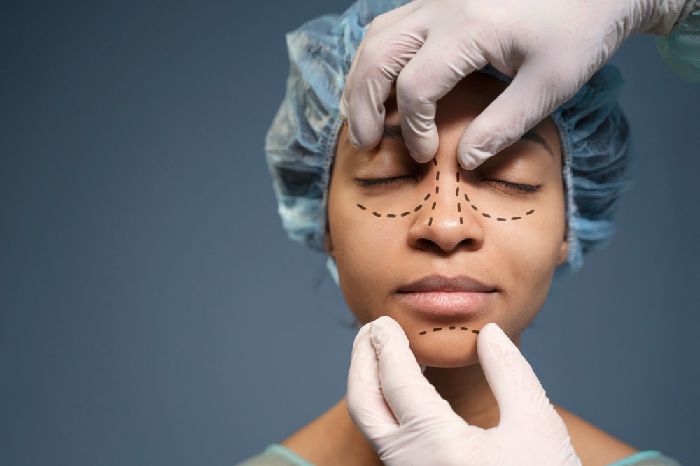
In rhinoplasty, osteotomy describes the controlled fracturing and repositioning of the nasal bones as part of reshaping the nose. It is performed to narrow a wide bony dorsum, close the open roof deformity that results from removing a hump, or correct a deviated bony nasal framework. Without osteotomy, changes to the cartilaginous lower two-thirds of the nose cannot be fully integrated with the bony upper third.
Three cut directions are typically combined in rhinoplasty osteotomy:
- Lateral osteotomies — run along the side walls of the nasal bones to allow inward movement
- Medial osteotomies — placed along the upper border of the nasal bones to free them from the nasal septum
- Transverse osteotomies — made across the upper nasal bones when additional mobilisation is needed
Periorbital swelling and bruising in the days following surgery are expected consequences of the procedure and typically resolve within ten to fourteen days.
Osteotomy Surgery at Medical Park
Medical Park Hospitals in Turkey perform osteotomy procedures across orthopedic surgery, oral and maxillofacial surgery, and plastic surgery, with specialist teams for each area operating within dedicated programmes.
Orthopedic osteotomies including high tibial osteotomy, tibial tubercle osteotomy, periacetabular osteotomy, and femoral osteotomy are performed within a joint preservation programme that provides a structured pathway for patients who are not yet appropriate candidates for joint replacement. Foot and ankle osteotomies — covering the full range of calcaneal, hallux, lesser metatarsal, and midfoot procedures — are performed by specialist foot and ankle surgeons.
Orthognathic jaw surgery is carried out by oral and maxillofacial surgeons working in coordination with orthodontic teams following three-dimensional surgical planning. Rhinoplasty osteotomy is available through Medical Park's plastic surgery department.
International patients seeking osteotomy surgery in Turkey benefit from access to specialist care, potentially shorter waiting times than in some healthcare systems, and lower overall treatment costs than in Western Europe and North America. Medical Park's international patient services team coordinates pre-operative assessment, surgical care, and post-operative follow-up, providing patients with a coordinated treatment pathway from initial consultation through recovery





 Makaleye Dön
 E-Result
 WhatsApp