


 TR
 EN
 RU
 AR
 FR


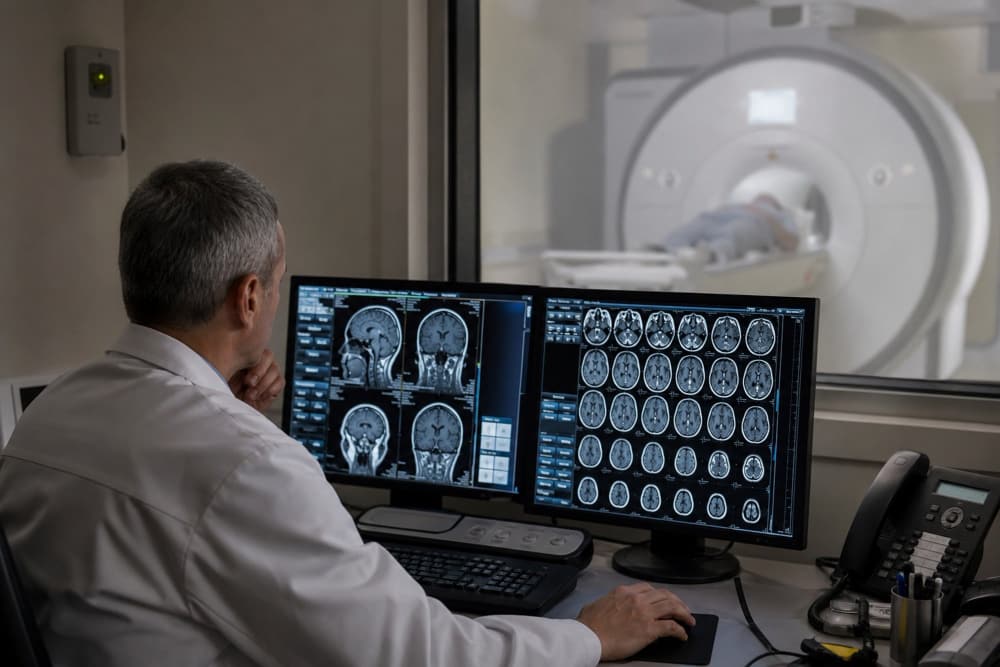

A SPECT scan, short for single-photon emission computerized tomography, is a nuclear medicine imaging test that helps doctors assess how organs and tissues function. Unlike imaging tests that primarily show the body's structure, SPECT imaging tracks a small amount of radioactive tracer as it moves through the body, providing information about blood flow and activity in specific tissues.
It is used in several areas of medicine, including heart disease evaluation, brain imaging, and the assessment of certain bone conditions. SPECT can also be combined with computed tomography (CT) to help locate abnormal activity more precisely.
Understanding what a SPECT scan can reveal, how the procedure works, and how its results are interpreted can help patients prepare for the test and understand its role in diagnosis and treatment planning.
This guide explains what a SPECT scan is used for, how the procedure works, how to prepare for the test, what its results may reveal, and how SPECT compares with PET scans.
What Is a SPECT Scan?
Single-photon emission computed tomography, also called single-photon emission computerized tomography, is a type of nuclear medicine imaging. It uses a radiotracer—a substance containing a small amount of radioactive material—to create three-dimensional images of activity inside the body.
The radiotracer is usually injected into a vein and travels through the bloodstream to the tissues being examined. A specialized camera detects the gamma rays released by the tracer, and a computer processes the signals into images. The resulting SPECT image can show how blood reaches an organ or how a particular tissue functions.
SPECT differs from conventional CT because the two tests provide different types of information:
SPECT imaging: Shows the distribution of a radiotracer and provides functional information, such as blood flow or activity in specific tissues.
CT imaging: Uses X-rays to produce detailed pictures of anatomical structures, including bones and organs.
Combined SPECT/CT: Brings functional SPECT images together with CT images to help identify the anatomical location of an abnormal finding.
SPECT does not provide a diagnosis on its own. Doctors interpret the images alongside symptoms, medical history, examination findings, laboratory tests, and other imaging results.
What Is a SPECT Scan Used For?
The purpose of a SPECT scan depends on the organ being examined and the radiotracer selected. It may help investigate a suspected condition, assess the extent of an abnormality, or evaluate how well a treatment has worked.
Common applications include:
Heart conditions: Assessing blood flow to the heart muscle, identifying areas that receive insufficient blood, and evaluating damage following a heart attack.
Neurological conditions: Examining blood flow or specific patterns of activity in the brain when investigating selected neurological disorders.
Bone conditions: Detecting abnormal bone activity associated with certain fractures, infections, or other bone disorders.
Infection and inflammation: Identifying areas of increased activity in selected cases when a specific nuclear medicine study is appropriate.
Certain cancer-related assessments: Helping locate particular abnormalities or evaluate specific conditions, depending on the radiotracer and clinical question.
SPECT is not the preferred test for every condition. A doctor selects the imaging method based on the information needed, the suspected diagnosis, and the advantages and limitations of available tests.
Brain SPECT Scan: What Does It Show?
A brain SPECT scan evaluates patterns of blood flow in the brain. Because brain activity is closely related to its blood supply, differences in tracer distribution can provide information about how different regions are functioning.
Brain SPECT imaging may be used in selected neurological assessments, including certain cases involving epilepsy, movement disorders, or other conditions that affect brain function. The exact role of the scan depends on the clinical question and the availability of other diagnostic methods.
A brain SPECT scan may help doctors:
Assess regional blood flow: Identify areas with relatively increased or decreased tracer uptake.
Support epilepsy evaluation: In selected patients, SPECT performed during or around a seizure may help identify a region associated with seizure activity as part of a broader assessment.
Investigate selected movement disorders: Functional imaging may provide additional information when evaluating certain neurological symptoms.
Contribute to complex diagnostic assessments: Findings may be considered alongside neurological examination, MRI, EEG, laboratory tests, or other investigations.
Does a Brain SPECT Scan Diagnose Alzheimer's Disease or Dementia?
A brain SPECT scan may show patterns of altered blood flow, but these patterns are not specific to a single disease. They cannot independently confirm Alzheimer's disease or distinguish every cause of memory loss, including frontotemporal dementia.
The diagnosis of dementia usually involves a detailed medical history, cognitive assessment, neurological examination, laboratory investigations, and structural brain imaging. Additional tests are selected according to the patient's symptoms and the suspected cause. Brain SPECT may be considered in particular clinical circumstances, but it is not a routine first-line test for every person with memory problems.
What Is a SPECT/CT Scan?
A SPECT/CT scan combines single-photon emission computerized tomography with computed tomography in one imaging system. The SPECT component shows where the radiotracer accumulates, while CT provides anatomical detail that helps doctors determine the location of the activity.
This combination can be useful when SPECT images alone do not clearly show which structure is responsible for an abnormal finding. For example, SPECT/CT may help localize areas of abnormal bone activity or clarify findings in selected endocrine, infection, or cardiac-related investigations.
Potential advantages of SPECT/CT include:
More precise localization: Functional findings can be matched to specific anatomical structures.
Improved interpretation: CT may help distinguish activity in a particular organ or bone from activity in nearby tissues.
Additional diagnostic information: Combining functional and anatomical images may help answer a specific clinical question more clearly than either technique alone.
SPECT/CT scanners are designed to acquire both sets of images during the same examination. However, the CT component uses X-rays and adds to the overall radiation exposure. Whether combined imaging is appropriate depends on the clinical indication; it is not necessary for every SPECT scan.
SPECT vs. PET Scans: What Is the Difference?
SPECT and positron emission tomography (PET) are both nuclear medicine imaging techniques. They use radiotracers to provide functional information, but they detect different types of radiation and use different imaging technologies. The most appropriate option depends on the condition being investigated, the tracer available, and the clinical information required.
| Feature | SPECT | PET |
|---|---|---|
| Full name | Single-photon emission computed tomography | Positron emission tomography |
| Imaging method | Detects gamma rays emitted by a radiotracer | Detects pairs of photons produced after positron annihilation |
| Common applications | Cardiac perfusion, selected brain studies, and bone imaging | Cancer imaging, selected neurological studies, and cardiac imaging |
| Image characteristics | Often lower sensitivity and spatial resolution than PET, depending on the system and tracer | Often offers higher sensitivity and spatial resolution, depending on the system and tracer |
| Combined imaging | Commonly performed as SPECT/CT | Commonly performed as PET/CT |
Which Is Better: SPECT or PET?
Neither test is universally better. PET may be preferred when a particular tracer can provide more sensitive information about metabolism or other biological processes. SPECT may be appropriate when established SPECT tracers can answer the clinical question, such as evaluating blood flow to the heart muscle.
Doctors consider several factors when choosing between SPECT and PET:
The condition being investigated and the information needed.
The availability and suitability of a tracer for the suspected condition.
The required image quality and the location of the suspected abnormality.
The patient's medical history and the need to minimize radiation exposure.
Local access to the necessary equipment and specialist interpretation.
The choice should be based on clinical need rather than the assumption that one type of scan is always more accurate.
How Is a SPECT Scan Performed?
The procedure varies according to the organ being examined and the radiotracer used. Some examinations require images to be taken after a waiting period, while cardiac studies may involve imaging at rest and during stress.
A typical SPECT scan involves the following steps:
Tracer administration: A healthcare professional usually injects the radiotracer into a vein. The injection itself may cause brief discomfort.
Waiting period: The tracer needs time to reach the tissues being examined. This may take minutes or several hours, depending on the study.
Positioning: You lie on an examination table, and the imaging team positions you so the camera can capture the required area.
Image acquisition: A gamma camera rotates around the body and detects signals from the tracer. You will generally need to remain still to reduce blurring.
Image processing: A computer reconstructs the collected data into cross-sectional and three-dimensional images. If SPECT/CT is required, CT images are also acquired.
The scan itself may take around 30 minutes or longer, but the total appointment can be several hours because of preparation, tracer uptake, and additional imaging. Cardiac studies may require more time if images are taken under different conditions.
SPECT is generally painless apart from the injection, and the camera does not touch or inject anything into the body during image acquisition. Tell the imaging team if you have difficulty lying still, experience discomfort, or feel anxious in enclosed spaces.
How to Prepare for a SPECT Scan
Preparation depends on the type of SPECT study. The imaging department will provide instructions specific to the tracer and the area being examined.
Before your appointment:
Ask about food and drink: Some scans require fasting, while others do not. Follow the instructions provided for your particular examination.
Discuss medications: Tell your doctor about prescription medicines, over-the-counter products, and supplements. Do not stop taking medication unless your healthcare team instructs you to do so.
Report pregnancy or breastfeeding: Tell the medical team if you are pregnant, think you may be pregnant, or are breastfeeding so they can assess the benefits, risks, and any necessary precautions.
Wear comfortable clothing: You may be asked to change into a hospital gown. Remove jewelry and other items that could interfere with imaging.
Mention relevant medical conditions: Inform the team about allergies, previous reactions to radiotracers, and any difficulty remaining still during the scan.
Follow cardiac test instructions: If the scan involves stress testing, you may receive additional instructions about caffeine, food, exercise, or medicines.
Following these instructions helps the team perform the examination safely and obtain images of suitable quality.
Are SPECT Scans Safe? Risks and Side Effects
SPECT scans use a small amount of radioactive material. The radiotracer is selected and administered in a controlled dose, and the potential benefit of the examination is weighed against the radiation exposure.
Possible considerations include:
Radiation exposure: SPECT involves ionizing radiation. The amount depends on the tracer, the examination, and whether CT is also performed.
Pregnancy: If you are pregnant or may be pregnant, the team must assess whether the scan is necessary and whether another test could provide the required information.
Breastfeeding: Some radiotracers may require temporary precautions or interruption of breastfeeding. Instructions depend on the specific tracer, so ask your nuclear medicine team for guidance.
Rare reactions: Adverse reactions to radiotracers are uncommon, but you should report any previous reaction to a nuclear medicine agent.
Stress-test risks: If the examination includes exercise or medication to stress the heart, the associated risks depend on your health and the method used.
Most people can resume their usual activities after a SPECT scan unless they have been given different instructions. The radiotracer loses radioactivity over time and is eliminated through the body's normal processes, although the time required varies by substance. Follow any specific hydration, bathroom, or contact precautions provided by the imaging team.
Understanding SPECT Scan Results
A nuclear medicine specialist or radiologist interprets the images and prepares a report for the doctor who requested the scan. The meaning of the findings depends on the organ examined, the radiotracer used, and the patient's symptoms and medical history.
A report may describe:
Expected tracer distribution: Activity appears consistent with the expected pattern for the examination.
Areas of increased uptake: A region shows more tracer accumulation than expected. The significance varies and may reflect several different processes.
Areas of reduced uptake: A region shows less tracer activity than expected, which may indicate reduced blood flow or another abnormality depending on the scan.
Limitations or unclear findings: Movement, technical factors, or overlapping activity may affect image interpretation and lead to a recommendation for further assessment.
An abnormal SPECT image does not automatically mean that a serious disease is present, and a normal result does not exclude every possible condition. The doctor may compare the findings with CT, MRI, laboratory tests, or other investigations before reaching a diagnosis or recommending treatment.
SPECT Scan in Turkey at Medical Park
Medical Park Hospitals provide SPECT scans at several hospital locations in Istanbul and other major cities in Turkey, making nuclear medicine imaging accessible to patients who require further investigation of organ function, blood flow, or abnormal activity in specific tissues. Expert Nuclear Medicine specialists interpret SPECT scan findings alongside the patient's symptoms, medical history, physical examination, and other diagnostic results to assess their clinical significance.
Depending on the medical concern, SPECT may be used to evaluate blood flow to the heart muscle, investigate selected neurological conditions, or assess certain bone abnormalities. When SPECT/CT is indicated, combining functional imaging with detailed anatomical images can help localize abnormal tracer uptake and provide additional information for diagnosis and treatment planning.
Following the scan, the next steps depend on the findings and the condition being investigated. Patients undergoing cardiac assessment may require further evaluation by Cardiology specialists, while those with neurological symptoms may need additional investigations coordinated with Neurology. The treating team determines whether further imaging, laboratory tests, or specialist assessments are needed and explains how the results inform subsequent care.
Medical Park’s International Patient Services Center helps international patients coordinate appointments, communicate with the healthcare team, and navigate the diagnostic process when traveling to Turkey for medical care.


 Makaleye Dön
 E-Result
 WhatsApp