


 TR
 EN
 RU
 AR
 FR


Stroke is a sudden disruption of blood supply to the brain that causes brain cells to die within minutes, making it a leading cause of death and long-term disability worldwide. Recognizing the warning signs early and seeking emergency treatment immediately are the most critical factors in limiting brain damage and improving outcomes. With advances in acute care, rehabilitation medicine, and stroke prevention, many patients achieve meaningful recovery and return to an independent life.
What is a Stroke?
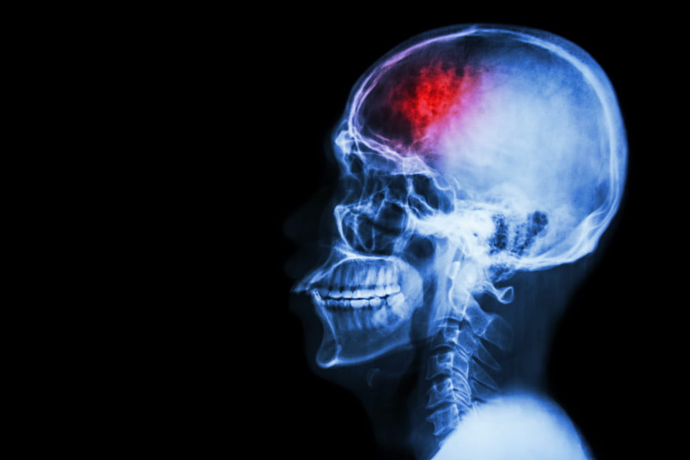
A brain stroke occurs when blood flow to part of the brain is cut off or severely reduced, depriving brain tissue of the oxygen and nutrients it needs to function. Within minutes of that disruption, brain cells begin to die—a process that can continue for hours if the underlying cause is not treated. The consequences range from temporary weakness or speech difficulties to permanent paralysis, cognitive impairment, or death, depending on which area of the brain is affected and how quickly treatment for stroke is received.
There are three main types of stroke: ischemic stroke, hemorrhagic stroke, and transient ischemic attack (TIA). Understanding the differences between these conditions is critical because their causes, management, and long-term outlooks vary significantly.
Ischemic Stroke
Ischemic stroke (also spelled ischaemic stroke) is by far the most common form, accounting for approximately 87 percent of all cases. It occurs when a blood clot or fatty deposit (plaque) blocks an artery supplying blood to the brain. There are two primary mechanisms:
Thrombotic Stroke: A blood clot forms directly inside an already narrowed cerebral artery, typically at the site of a ruptured or eroded atherosclerotic plaque. This localized buildup blocks blood flow to the downstream brain tissue, immediately causing an ischemic stroke in the specific area supplied by that vessel. It is the most common subtype of ischemic stroke, frequently linked to chronic vascular inflammation, diabetes, and uncontrolled hypertension.
Embolic Stroke: A clot forms elsewhere in the body—most often the heart, a large neck artery, or as a consequence of systemic clotting issues like a deep vein thrombosis that travels—and moves through the bloodstream until it lodges in a smaller brain vessel. While clots from the venous system typically travel to the lungs to cause a pulmonary embolism, certain cardiac anatomical variations (such as a patent foramen ovale) can allow a venous clot to cross into the arterial system and travel to the brain, causing an embolic stroke.
Hemorrhagic Stroke
Hemorrhagic stroke occurs when a blood vessel in or around the brain ruptures, flooding surrounding tissue with blood. This type is broadly divided into two categories:
Intracerebral Hemorrhage: Bleeding occurs directly within the brain tissue itself, most commonly due to chronic high blood pressure weakening the arterial walls.
Subarachnoid Hemorrhage: Blood leaks into the space between the brain and its outer membrane, often triggered by a ruptured brain aneurysm.
While hemorrhagic variants are less common, they carry a higher risk of severe stroke complications and mortality because the pooling blood creates mechanical pressure that damages brain cells far beyond the original rupture site.
Transient Ischemic Attack (TIA)
A transient ischemic attack —widely known as a ministroke or TIA—produces stroke-like symptoms that resolve completely within 24 hours, usually within minutes. Although a ministroke causes no permanent brain injury, it is a powerful warning sign that a full brain stroke may follow. The risk of an acute event in the days immediately following a TIA is among the highest of any cardiovascular condition, meaning it requires the same urgent investigation and risk-factor management as a completed stroke.
Recognizing the Critical Signs of Stroke
For years, the traditional FAST acronym was the primary public screening tool for catching common acute stroke indicators. However, contemporary clinical practice has moved toward the more comprehensive BE-FAST framework, which expands on standard protocols by catching critical early neurological indicators associated with strokes affecting the posterior blood supply of the brain.
The Shift From FAST to BE-FAST: Why the Screening Framework Expanded?
While standard screening protocols remain useful, incorporating the extra diagnostic categories increases early identification rates significantly. Data indicates that using the updated clinical tool helps medical teams and bystanders successfully recognize up to 95% of ischemic stroke cases, a major improvement over the 85% detection rate of the older method.
B - Balance Impairment: A sudden, unexplained loss of coordination, instability, or acute difficulty walking normally without a clear physical cause.
E - Eyesight Changes: Sudden visual disturbances in one or both eyes, including unexplained double vision, blurred vision, or a complete blind spot in part of the visual field.
F - Facial Drooping: Pronounced weakness, numbness, or asymmetry on one side of the face, which becomes most visible when a person attempts to smile or show their teeth.
A - Arm Weakness: Sudden loss of motor strength or severe numbness in a single limb, causing the arm to drift downward when both arms are extended straight out.
S - Speech Disruptions: Slurred articulation, garbled word choice, a complete inability to find words, or a sudden failure to comprehend spoken language.
T - Time to Act: Every minute of untreated brain ischemia compromises critical neural tissue; immediate emergency services must be contacted the moment any of these warning signs appear.
Atypical Manifestations and Differences in Women
Beyond these core structural deficits, a cerebrovascular accident can also manifest through severe, instantaneous headaches with no known cause. Furthermore, women frequently present with less typical clinical signs, including sudden nausea, hiccups, chest discomfort, shortness of breath, or generalized body fatigue, which can dangerously delay life-saving emergency intervention if not caught right away.
Stroke Causes and Risk Factors
The underlying causes of stroke depend heavily on the specific type. Ischemic events are most often caused by atherosclerosis—the gradual build-up of fatty plaques inside arterial walls—combined with blood clotting disorders or cardiac conditions like atrial fibrillation. Conversely, hemorrhagic strokes are most commonly caused by uncontrolled high blood pressure that weakens arterial walls over time, though aneurysms and anticoagulant medications also play important roles.
Key modifiable stroke risk factors include:
High Blood Pressure (Hypertension): The single most important preventable risk factor for all stroke types.
Atrial Fibrillation: An irregular heart rhythm that allows blood clots to form in the heart and travel to the brain.
Type 2 Diabetes: A chronic condition that accelerates atherosclerosis and increases clotting tendencies.
High LDL Cholesterol: Contributes directly to plaque formation in cerebral and carotid arteries.
Cigarette Smoking: Damages blood vessel walls, narrows arteries, and accelerates clot formation.
Obesity and Physical Inactivity: Compounds vascular risk factors through multiple systemic mechanisms.
Excessive Alcohol Consumption: Raises baseline blood pressure and increases hemorrhagic risk.
Non-modifiable risk factors include increasing age, male sex (though women have a higher lifetime risk and worse overall outcomes), family history of stroke, and a prior history of a stroke or TIA.
Diagnosing Stroke
An accurate stroke diagnosis begins the moment a patient arrives in the emergency department. Time is the most critical variable: every minute of delayed diagnosis represents a minute of ongoing brain damage. The diagnostic process combines a rapid neurological assessment with urgent brain imaging to determine whether the stroke is ischemic or hemorrhagic—a distinction that directly determines which treatments are safe to administer.
Non-Contrast CT Scanning: The first-line imaging test because it can be performed within minutes and reliably excludes intracranial hemorrhage.
MRI Diffusion-Weighted Imaging: Used if the initial CT is normal but an ischemic event is still highly suspected, detecting early ischemic changes that standard CT may miss.
CT/MR Angiography: Visualizes the cerebral and neck arteries to identify the exact location and extent of any blockage or vascular malformation.
Laboratory & Cardiac Tests: Includes full blood counts, coagulation screens, blood glucose monitoring, electrocardiograms (ECG) to detect atrial fibrillation, and echocardiography to check for cardiac clots.
Carotid Ultrasound: Often used for TIA patients to evaluate the degree of narrowing in the carotid arteries to determine if urgent intervention is required.
Stroke Treatment Options
Effective treatment for stroke depends on rapid diagnosis, correct classification of the stroke type, and access to specialist care. The window for the most impactful interventions is measured in hours, which is why acute episodes are treated as medical emergencies in specialist stroke units equipped to deliver around-the-clock care.
Emergency intervention for ischemic stroke revolves around restoring blood flow:
Intravenous Thrombolysis: Administration of stroke medicines like alteplase (rtPA) to dissolve the clot, which must be given within 4.5 hours of symptom onset.
Mechanical Thrombectomy: A minimally invasive procedure where a catheter is passed through the arterial system to physically retrieve a clot in a major cerebral vessel. This can extend up to 24 hours in selected patients.
Acute Antiplatelet Therapy: Initial use of medicines like aspirin to prevent early recurrence.
Management for hemorrhagic stroke focuses on stabilization and pressure management:
Bleeding & Pressure Control: Rapid regulation of systemic blood pressure and reducing intracranial pressure inside the skull.
Anticoagulant Reversal: Immediate administration of targeted reversal agents for patients on blood thinners.
Neurosurgical Intervention: Surgical drainage, aneurysm clipping, or endovascular coiling for large hematomas causing mass effect or ruptured vascular structures.
Stroke Recovery and Rehabilitation
Stroke recovery is a progressive process, not a single event. After the acute phase, the brain begins a period of neuroplasticity—its ability to reorganize and form new connections to compensate for damaged areas. Most neurological recovery occurs in the first three to six months after a stroke, but meaningful improvement can continue for years with consistent therapy.
A comprehensive stroke rehabilitation program is multidisciplinary, addressing the physical, cognitive, communicative, and psychological consequences of brain injury:
Physiotherapy: Targets motor function, balance, and gait, using task-specific practice and progressive exercise to rebuild movement patterns.
Occupational Therapy: Helps patients relearn the activities of daily living—dressing, cooking, personal care—and recommends adaptive equipment to support independence at home.
Speech and Language Therapy: Addresses aphasia (difficulty understanding or producing language) and dysphagia (swallowing difficulties), both of which are common after a stroke.
Neuropsychological Support: Manages post-stroke depression, which affects approximately one in three stroke survivors, while addressing deficits in attention, memory, and executive function.
Many specialist centers also offer advanced options like robotic-assisted therapy, non-invasive brain stimulation, and virtual reality programs to augment conventional rehabilitation and accelerate recovery outcomes.
Preventing Future Strokes
Effective stroke prevention is the most impactful tool in cerebrovascular medicine. Because the majority of strokes are caused by modifiable risk factors, systematic management of those factors can reduce risk by more than 80 percent. Prevention strategies are broadly divided into primary prevention—reducing first stroke risk—and secondary prevention, which applies to survivors seeking to prevent recurrence.
Medical prevention strategies include:
Antihypertensive Therapy: The most important pharmacological intervention in stroke prevention, regardless of stroke type.
Anticoagulation Therapy: Typically with direct oral anticoagulants for patients with atrial fibrillation to dramatically reduce embolic risk.
Statin Therapy: Lowers LDL cholesterol and stabilizes arterial plaques for broader vascular protection.
Antiplatelet Agents: Medications such as aspirin or clopidogrel used after an ischemic stroke to prevent recurrent clot formation.
Carotid Interventions: Carotid endarterectomy or stenting for patients with significant carotid artery stenosis to clear the obstruction.
Lifestyle modifications remain fundamental to stroke prevention and complement medical therapy at every stage. Adopting a diet rich in vegetables, legumes, whole grains, and olive oil reduces cardiovascular risk through multiple pathways. Regular aerobic exercise—at least 150 minutes of moderate-intensity activity per week—lowers blood pressure, improves insulin sensitivity, and reduces overall vascular risk. Smoking cessation is among the most powerful individual interventions available: the excess stroke risk associated with smoking falls sharply within months of quitting and approaches non-smoker levels within five years.
Living with Stroke
Life after stroke presents challenges that extend well beyond the immediate medical recovery. Many survivors live with lasting stroke complications including partial paralysis, fatigue, cognitive changes, emotional dysregulation, and chronic pain. Adapting to these changes requires long-term support from healthcare professionals, family members, and peer networks. Specialist stroke nurses and community rehabilitation teams play an essential role in bridging the gap between hospital discharge and independent life at home.
Fatigue is one of the most pervasive and least understood sequelae of stroke, affecting more than half of survivors at some point and lasting years in many cases. Unlike ordinary tiredness, post-stroke fatigue is disproportionate to physical exertion and does not fully resolve with rest. Pacing strategies, energy management techniques, and — where appropriate — pharmacological support can help patients manage fatigue and maintain functional activity levels.
Returning to driving, work, and social activities depends on the nature and severity of residual deficits and varies considerably between individuals. Formal neuropsychological assessment can clarify cognitive fitness for specific tasks. Support groups — whether in-person or online — provide peer connections that reduce social isolation and help survivors and caregivers share practical coping strategies. Carers themselves are at risk of burnout and benefit from dedicated psychological support and respite care provision.
Acute Stroke Treatment and Post-Stroke Rehabilitation at Medical Park
International patients seeking acute stroke treatment or post-stroke rehabilitation at Medical Park Hospitals Group benefit from a fully integrated Stroke Center that brings together top-tier neurology, interventional radiology, brain and nerve surgery specialists and multidisciplinary rehabilitation under one roof.
Medical Park’s Stroke Centers operate 24 hours a day, seven days a week, and are equipped with advanced neuroimaging systems capable of performing CT angiography and MRI perfusion studies at any hour. Stroke medicines including intravenous thrombolytics and anticoagulants are available immediately on confirmed diagnosis, and mechanical thrombectomy for large vessel occlusion is performed by experienced interventional teams with direct access to hybrid operating theatres.
For patients transitioning from acute care into recovery, Medical Park’s neurorehabilitation centres deliver structured inpatient and outpatient programmes staffed by physiotherapists, occupational therapists, speech and language therapists, neuropsychologists, and specialist stroke nurses. Robotic technology and non-invasive deep brain stimulation protocols supplement conventional therapy to maximise neurological recovery.




 Makaleye Dön
 E-Result
 WhatsApp