


 TR
 EN
 RU
 AR
 FR


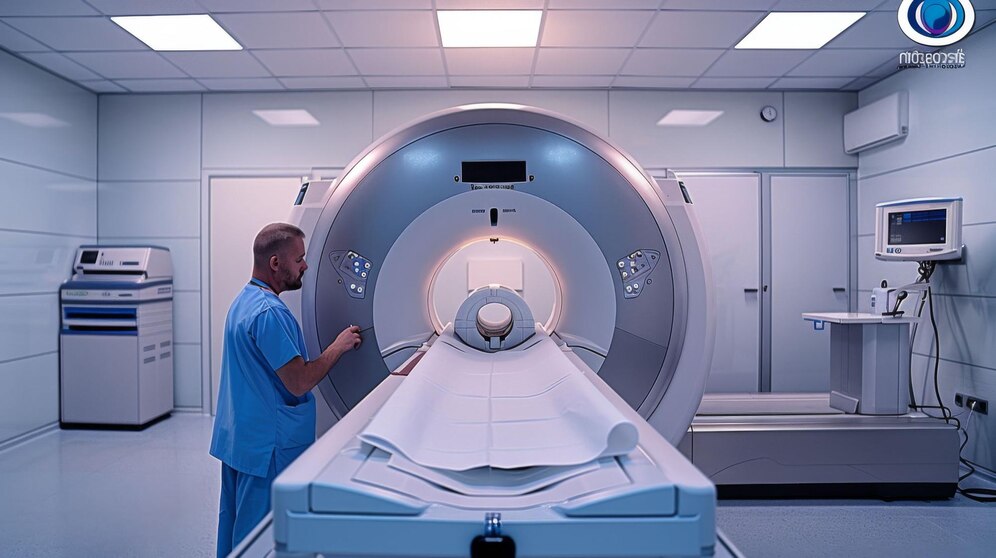

Nuclear Medicine is a specialized field of medicine that uses small amounts of radioactive substances, called radiotracers, to visualize, diagnose, and monitor the function of organs and tissues in the body. Unlike traditional imaging techniques that show only anatomical structures, nuclear medicine provides unique information about cellular activity, blood flow, and metabolic processes, allowing early detection of diseases, accurate assessment of organ function, and precise evaluation of treatment effectiveness.
Conditions Diagnosed
Heart Diseases: in collaboration with Cardiology and Cardiovascular Surgery
Thyroid and Endocrine System Diseases
Bone Diseases: Bone Scan, Total Body Bone Scan, 3-phase Bone Scan, Leukocyte Labeling
Renal and Genital System Diseases: Dynamic and Static Renal Scans, Vesicoureteral Reflux (VUR) Scan
Gastrointestinal System Diseases: disorders of the stomach, digestive tract, liver, and bile ducts
Cancer
Other Conditions
Specialized Scans
Lung Perfusion/Ventilation Scan for pulmonary embolism
Lacrimal Duct Scan for lacrimal duct obstructions
Parotid Gland Scan for parotid gland diseases
Ga-67 or Labeled Leukocyte Scan for infections, abscesses, and idiopathic fever
Labeled Leukocyte Scan for diabetic foot evaluation
Bone Marrow Scan
PET/CT: Advanced Imaging Technology
PET/CT combines two advanced imaging techniques to provide detailed information for diagnosis, treatment, and follow-up, particularly in cancer care.
PET (Positron Emission Tomography): reveals cellular function and metabolism
CT (Computed Tomography): provides precise anatomical details
Together, PET/CT visualizes both metabolism and anatomy in 3D during a single session, enabling accurate assessment and treatment planning. Our PET/CT system at the cancer hospital can detect even the smallest tumors.
PET vs PET/CT
PET alone can detect tumor metabolism but cannot accurately locate lesions without CT guidance.
PET/CT scans are faster and designed with a spacious structure for patient comfort, including those with claustrophobia.
Uses of PET/CT
Cancer diagnosis, staging, and evaluation of treatment response
Cardiology: determining viable cardiac tissue after myocardial infarction
Neurology: precise localization of epileptic foci
Early detection of neurodegenerative diseases such as Alzheimer’s
Role in Radiotherapy
PET/CT-guided radiotherapy has become a global standard
Helps precisely target tumor tissue while minimizing damage to surrounding healthy tissue
How PET/CT Scans Are Performed
A radioactive contrast agent is injected
Patient rests 45–60 minutes to allow uptake by cancerous cells
Scanning is performed while the patient lies in a supine position for 35–45 minutes
Advantages of PET/CT
Early and precise cancer detection
Identification of recurrences at an earlier stage
High-quality, detailed imaging
Increased patient comfort due to shorter scan duration
Enhanced effectiveness of radiotherapy, conserving healthy tissue
This content was developed by the Medical Park Editorial Board and is intended for informational purposes only. It does not replace professional medical advice, diagnosis, or treatment. Please consult your healthcare provider for personalized care.


 E-Result