


 TR
 EN
 RU
 AR
 FR


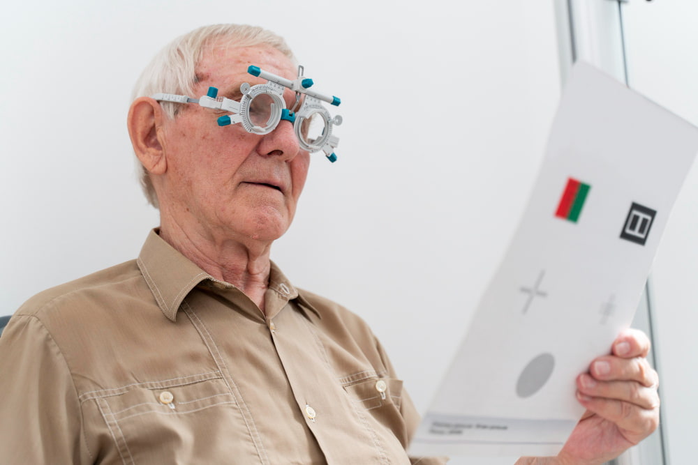


Optic atrophy is a condition characterized by damage to the optic nerve, the crucial pathway that carries visual information from your eye to your brain. In simple terms, “atrophy” means wasting away, so optic atrophy refers to the deterioration of the optic nerve fibers. When these nerve fibers are damaged, they cannot transmit visual signals effectively, which can result in vision loss.
Also called optic nerve atrophy, this condition represents the end stage of various causes of optic nerve damage. It is important to understand that optic atrophy itself is not a disease but a sign that the optic nerve has been compromised due to underlying conditions. Since the optic nerve is responsible for conveying retinal information to the brain, any damage can have serious implications for vision, including permanent blindness.
The optic nerve consists of approximately 1.2 million axons from retinal ganglion cells. These axons are myelinated by oligodendrocytes, which do not regenerate once damaged. As a result, optic nerve atrophy is usually irreversible, making early detection and management critical.
Causes of Optic Atrophy
Optic atrophy can occur due to a wide range of factors, all of which interfere with the optic nerve’s ability to transmit signals. The most common causes of optic atrophy include:
Vascular/Ischemic Causes: Reduced blood flow to the optic nerve is the most frequent cause of optic atrophy. Conditions such as nonarteritic ischemic optic neuropathy can result in permanent nerve damage.
Congenital or Hereditary Causes: Some individuals are born with conditions that predispose them to optic nerve degeneration. For instance, Leber hereditary optic neuropathy typically causes vision loss first in one eye and then the other.
Compression: Pressure on the optic nerve can occur externally or internally. Tumors, increased intraocular pressure from glaucoma, or structural abnormalities can compress the nerve, leading to atrophy.
Inflammation: Inflammatory conditions, such as optic neuritis or swelling due to hydrocephalus (fluid accumulation in the brain), can damage the optic nerve.
Infections: Certain infections, including syphilis, measles, tuberculosis, mumps, chickenpox, Lyme disease, aspergillosis, and cryptococcosis, may affect the optic nerve.
Trauma: Eye injuries from accidents, sports, or physical altercations can directly damage optic nerve fibers.
Retinal Diseases: Conditions like diabetic retinopathy or retinal vein occlusion can indirectly damage the optic nerve.
Toxins, Nutritional Deficiencies, or Medications: Exposure to certain toxins, lack of essential nutrients, or side effects of medications (e.g., ethambutol, amiodarone) can contribute to optic nerve atrophy. These causes often affect both eyes simultaneously.
It is important to note that optic atrophy is not contagious, even though infections may trigger the condition.
Symptoms of Optic Atrophy
The symptoms of optic nerve atrophy are primarily related to vision changes. Common optic atrophy symptoms include:
Blurred vision or decreased visual sharpness
Reduced peripheral vision
Difficulties distinguishing colors or color vision changes
Loss of contrast sensitivity
Depending on the underlying cause, additional symptoms may occur. For instance, inflammation may cause eye pain, while glaucoma-related optic atrophy may be painless but progressive.
Because the optic nerve transmits information from the retina to the brain, any compromise in this pathway will gradually affect the clarity and field of vision. Severe cases can result in legal blindness or complete loss of light perception.
Diagnosing Optic Atrophy
Early and accurate diagnosis is essential for managing optic atrophy and preventing further vision loss. Eye care specialists use a combination of medical history, physical examination, and diagnostic tests to assess optic nerve damage.
Medical History
Your ophthalmologist will ask detailed questions about:
The onset and progression of vision loss
Any previous eye conditions or injuries
Medications, supplements, and lifestyle factors
Family history of eye or hereditary diseases
This helps identify potential causes and guides further investigations.
Physical Examination
A thorough eye exam includes:
Assessment of visual acuity, color, and contrast vision
Visual field testing
Intraocular pressure measurement
Pupillary reflex evaluation for afferent defects
Fundoscopy to examine the optic disc
Characteristic signs of optic atrophy on examination include a pale optic disc, sharp disc margins, and thinning of the retinal nerve fiber layer.
Diagnostic Tests
Additional tests help confirm the diagnosis and identify the underlying cause:
Optical Coherence Tomography (OCT): Measures the thickness of the nerve fiber layer and retinal ganglion cells.
Visual Field Tests: Detect areas of vision loss and help localize the affected part of the optic nerve.
Magnetic Resonance Imaging (MRI) or CT Scans: Detect tumors, structural abnormalities, or inflammation along the optic pathway.
Fluorescein Angiography: Evaluates blood flow to the optic nerve and retina.
Ultrasound: Useful for detecting structural eye changes.
Blood Tests: Screen for infections, vitamin deficiencies, autoimmune conditions, or systemic diseases contributing to optic nerve damage.
Electrophysiological Testing (ERG, VEP): Helps differentiate optic nerve pathology from retinal disease.
Differential Diagnosis
Optic atrophy may sometimes be confused with:
Optic nerve hypoplasia
Myelinated nerve fibers
Tilted optic disc
Myopic crescent
Careful examination and diagnostic testing usually allow clinicians to distinguish these conditions from true optic nerve atrophy.
Treatment and Management of Optic Atrophy
Currently, there is no cure for optic atrophy. Once optic nerve fibers are damaged, they cannot regenerate. Treatment focuses on addressing the underlying cause and preventing further damage.
Managing Underlying Causes
Tumors or Compressive Lesions: Surgical removal or treatment of tumors can relieve pressure on the optic nerve.
Glaucoma: Reducing intraocular pressure can slow the progression of optic nerve damage.
Inflammation: Treating conditions like optic neuritis or systemic inflammatory diseases may prevent further damage.
Vascular Conditions: Managing diabetes, hypertension, or other vascular risk factors can help reduce additional nerve injury.
Infections and Toxins: Prompt treatment of infections and avoidance of toxic agents are essential.
Vision Support
For individuals with irreversible optic nerve damage, low-vision aids may improve quality of life:
Special lenses or magnifying glasses
Text-to-speech or screen reading software
Adaptive lighting and contrast-enhancing devices
Early detection remains key to preserving remaining vision, and routine eye exams play a crucial role in monitoring at-risk patients.
Prevention of Optic Atrophy
While optic atrophy itself cannot usually be prevented, certain measures may reduce the risk of optic nerve damage:
Wearing protective eyewear to prevent eye injuries
Receiving recommended vaccinations to avoid infections
Practicing safe sex to prevent sexually transmitted infections
Managing chronic diseases such as diabetes or hypertension
Regular eye check-ups to detect conditions like glaucoma early
Prompt attention to vision changes can help prevent progression and preserve remaining sight.
Living With Optic Atrophy
Regular monitoring and early intervention are essential for individuals with optic atrophy. You should see a healthcare provider if you experience:
Blurry or reduced vision
Difficulty with color perception
Loss of peripheral vision
Sudden loss of vision or severe eye pain (seek immediate medical attention)
Although optic atrophy cannot be reversed, stabilizing the condition and protecting remaining vision are achievable with timely medical care and supportive strategies. You can consult with our experienced ophthalmology specialists at Medical Park for a comprehensive evaluation and personalized care for optic atrophy and other eye conditions.




 Makaleye Dön
 E-Result
 WhatsApp