


 TR
 EN
 RU
 AR
 FR


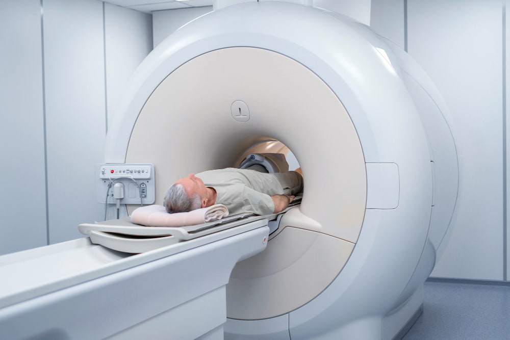

A PET/CT scan is an advanced imaging method that combines positron emission tomography (PET) and computed tomography (CT) in a single examination. This hybrid approach allows clinicians to evaluate both metabolic activity and anatomical structure at the same time, making PET/CT one of the most powerful tools in modern medical imaging—particularly in cancer care.
This guide explains how PET/CT works, its clinical uses, how to prepare, and what to expect during the procedure.
What Is a PET/CT Scan?
PET/CT is performed using a single integrated scanner that acquires both PET and CT images during the same session. The CT component provides detailed anatomical information, while the PET component detects metabolic activity using a radioactive tracer. These datasets are digitally fused to create highly precise, three-dimensional images.
The CT scan uses X-rays to show the size, shape, and location of organs and tissues. The PET scan, by contrast, highlights how tissues are functioning. A small amount of tracer is injected into the bloodstream, and areas with increased metabolic activity appear more prominent on imaging.
By combining these two technologies, PET/CT can show not only where an abnormality is located but also whether it is biologically active—an advantage that neither PET nor CT can provide alone.
The Role of PET
A PET scan on its own focuses on metabolic processes in the body. While it is valuable in certain neurological and cardiac applications, it is most commonly used today as part of PET/CT, where anatomical localisation significantly improves diagnostic accuracy. For this reason, PET-only imaging plays a more limited role in routine clinical practice compared to PET/CT.
Fluorodeoxyglucose (FDG): The Standard Tracer
Most PET/CT scans use fluorodeoxyglucose (FDG), a glucose-based radioactive tracer. Because active cells consume more glucose, FDG accumulates in areas of high metabolic activity.
Cancer cells, infections, and inflammatory processes often show increased FDG uptake. This makes FDG PET/CT highly effective in identifying abnormal tissue, although results must always be interpreted within the broader clinical context.
What Is PET/CT Used For?
PET/CT is used across several medical specialties, but its primary role is in oncology.
PET/CT in Oncology
PET/CT is considered a standard imaging method in cancer care. It supports:
Cancer detection: Identifying metabolically active tumors, sometimes before structural changes are visible.
Staging: Determining whether cancer has spread to lymph nodes or distant organs.
Treatment planning: Helping guide radiotherapy by defining the exact tumor boundaries.
Response assessment: Evaluating how well a tumor responds to chemotherapy or radiation.
Recurrence detection: Identifying returning disease at an early stage.
An important advancement is PSMA PET/CT in prostate cancer. This technique uses a targeted tracer to detect prostate cancer cells with high sensitivity, even at low PSA levels.
Cardiac PET/CT
In cardiology, PET/CT is used to assess blood flow and heart muscle viability. The CT component in these cases is typically low-dose and used mainly to improve PET data accuracy.
The most common application is myocardial perfusion imaging, which evaluates blood supply to the heart muscle under rest and stress conditions. This helps determine whether procedures such as stenting or bypass surgery are needed.
Neurological Applications
Neurological PET imaging is more specialized and often combined with MRI rather than CT. When PET/CT is used in brain imaging, the CT component generally serves a supportive role.
PET can help identify patterns of brain metabolism associated with conditions such as Alzheimer’s disease, epilepsy, and movement disorders. However, these applications are more selective compared to the widespread use of PET/CT in oncology.

PET/CT vs CT Alone
CT scans provide detailed structural information but cannot always distinguish between active disease and inactive tissue. PET/CT adds metabolic insight, allowing clinicians to determine whether a lesion is biologically active.
For example, an enlarged lymph node on CT may be benign, while a small but metabolically active area on PET/CT may indicate early disease. This combination improves diagnostic accuracy and supports better treatment decisions.
How to Prepare for a PET/CT Scan
Proper preparation is essential for accurate results.
Diet and fasting:
Do not eat for at least 4–6 hours before the scan.
Avoid sugar, caffeine, and alcohol.
Drink water freely.
Blood sugar control is especially important. Patients with diabetes will receive individualized instructions.
Physical activity:
Avoid strenuous exercise for 24 hours before the scan, as it can affect tracer distribution.
Medications:
Inform your doctor about all medications.
Some drugs, particularly diabetes medications and those related to cardiac stress testing, may need adjustment.
What Happens During the PET/CT Procedure?
A small amount of radioactive tracer is injected intravenously. You will rest quietly for about 45–60 minutes while the tracer distributes in your body.
During the scan:
You lie on a motorized table that moves through the scanner.
The CT scan is performed first and takes only seconds.
The PET scan follows and takes approximately 20–40 minutes.
The procedure is painless, and you can return to normal activities afterward. Drinking fluids helps eliminate the tracer from your body.
Safety and Side Effects of PET/CT
PET/CT is generally safe and well tolerated.
Radiation exposure is low and considered acceptable for diagnostic purposes.
Side effects are rare and may include mild discomfort at the injection site.
Allergic reactions are extremely uncommon.
As a precaution, patients are usually advised to limit close contact with pregnant women and young children for a few hours after the scan. Pregnant or breastfeeding patients should inform their doctor in advance.
PET/CT at Medical Park
Medical Park provides PET/CT and PET/MRI imaging as part of an integrated, multidisciplinary approach to diagnosis and treatment planning. Nuclear medicine physicians, radiologists, oncologists, cardiologists, and neurologists collaborate to ensure that every scan is reported in the context of the patient's full clinical history and that findings translate into clear, timely management recommendations.
Oncology patients benefit from PET/CT services that cover the entire care pathway, from initial staging through treatment response assessment and long-term disease surveillance. Patients with cardiac disease have access to dedicated myocardial perfusion and viability imaging protocols. Neurological PET investigations, including FDG and amyloid imaging for the evaluation of dementia and other neurodegenerative conditions, are available within the same multidisciplinary framework.
International patients receive coordinated support throughout their visit, including preparation guidance, assistance with prior imaging review, interpreter services, and seamless referral to the relevant specialist following results.