


 TR
 EN
 RU
 AR
 FR


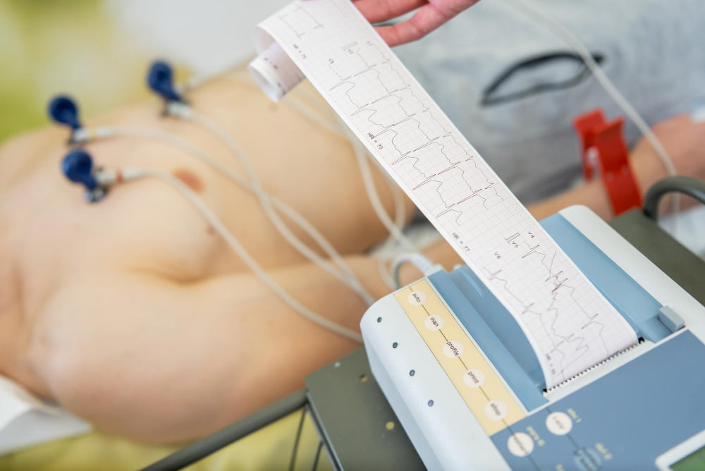

An electrocardiogram (EKG) is a quick, noninvasive test that records the electrical activity of the heart. An EKG test provides information about heart rate, rhythm, and how electrical signals travel through the heart. It is commonly used to investigate symptoms such as chest pain, palpitations, dizziness, and shortness of breath and to evaluate a range of cardiovascular conditions.
Because an EKG is painless, widely available, and takes only a few minutes, it is often one of the first tests performed when a heart problem is suspected. The results can help doctors determine whether further cardiac testing or treatment is needed.
What Is an Electrocardiogram (EKG)?
An electrocardiogram records the electrical signals produced by the heart during each heartbeat. Every heartbeat begins with an electrical impulse that travels through specialized pathways in the heart, causing the heart muscle to contract in a coordinated sequence. An electrocardiograph detects these signals and displays them as waves and lines on a screen or printed tracing.
The terms electrocardiogram, ECG, and EKG generally refer to the same test. "EKG" comes from the German spelling of the word and is commonly used in clinical settings.
An EKG can provide information about:
- Heart rate and rhythm
- The timing and direction of electrical impulses
- Electrical conduction through the heart
- Certain abnormal heart rhythms (arrhythmias)
- Premature heartbeats, including premature ventricular contractions
- Changes that may indicate reduced blood flow to the heart
- Patterns associated with a current or previous heart attack (myocardial infarction)
- Some changes related to enlargement or strain of the heart chambers
An EKG cannot evaluate every aspect of cardiovascular health. Depending on the patient's symptoms and findings, a doctor may recommend an echocardiogram, blood tests, heart rhythm monitoring, or other examinations.
How Does an EKG Test Work?
During an EKG test, small adhesive electrodes are placed on specific areas of the chest, arms, and legs. These electrodes detect the electrical signals generated by the heart and transmit them to the electrocardiograph, which converts them into a visual tracing.
The patient usually lies still on an examination table while the recording is made. A standard resting EKG generally takes only a few minutes. It does not involve needles or electrical stimulation, and the test does not send electricity into the body.
For accurate results, the skin may be cleaned before the electrodes are attached. Patients may also be asked to avoid applying creams or oils to the skin beforehand because these can interfere with electrode contact. Remaining still and relaxed during the recording can also help produce a clear tracing.
A standard EKG provides a snapshot of the heart's electrical activity at a particular moment. If symptoms occur intermittently, such as occasional palpitations or episodes of an irregular heartbeat, a doctor may recommend longer-term monitoring with a Holter monitor or another ambulatory heart monitor.
What Does an EKG Show?
An EKG shows how electrical activity moves through the heart and whether this activity follows an expected pattern. Doctors examine the waves, intervals, and segments of the tracing to assess the heart's rate, rhythm, and electrical conduction.
The main components of a normal EKG correspond to different stages of the heartbeat. The P wave represents electrical activation of the atria. The QRS complex represents ventricular depolarization, when the ventricles are electrically activated before they contract. The T wave represents ventricular repolarization, when the ventricles recover electrically in preparation for the next heartbeat.
Changes in these patterns can provide clues about a variety of cardiovascular problems. An EKG may help identify abnormal heart rhythms, including atrial fibrillation, as well as electrical conduction disorders, premature beats, or changes associated with reduced blood flow to the heart. It can also provide evidence of a current or previous myocardial infarction (heart attack) and may show changes related to enlargement or strain of the heart chambers.
An EKG may also reveal indirect changes associated with some forms of valve disease, such as mitral valve disease, although it cannot directly show the heart valves. Further testing may therefore be necessary to determine the underlying cause of an abnormal finding.
What Does a Normal EKG Look Like?
A normal EKG generally shows a regular heart rhythm, an appropriate heart rate, and electrical waves and intervals that fall within expected ranges. The electrical impulse should travel through the heart in an organized sequence, producing the characteristic P waves, QRS complexes, and T waves.
There is not one identical tracing that represents a normal EKG for everyone. Heart rate and certain aspects of the tracing can vary depending on factors such as age, physical condition, medications, and other individual characteristics.
For this reason, an EKG should be interpreted by a healthcare professional rather than judged by comparing the tracing with an example found online. Doctors consider the patient's symptoms and medical history as well as previous EKGs and other test results when determining whether a finding is clinically significant.
What Is an Abnormal EKG?
An abnormal EKG indicates that the heart's electrical activity differs from the pattern a doctor would typically expect. The significance of the finding can vary considerably. Some changes may be associated with an underlying heart condition, while others may result from temporary factors, medications, or individual variations in the way the heart's electrical activity appears on the tracing.
An abnormal result does not, by itself, establish a diagnosis or indicate how serious a condition may be. Doctors consider the specific pattern on the EKG together with the patient's symptoms, medical history, physical examination, and any previous test results.
If an EKG is abnormal, the next step depends on the nature of the finding and the patient's overall clinical picture. A doctor may repeat the EKG, compare it with an earlier tracing, or recommend additional tests such as an echocardiogram, blood tests, stress testing, or longer-term heart rhythm monitoring. This further evaluation helps determine whether the finding requires treatment or simply continued observation.
Some EKG findings may also prompt further evaluation for conditions such as congenital heart disease, particularly when the electrical pattern suggests an underlying structural abnormality. Additional tests, such as an echocardiogram, may then be used to assess the heart in greater detail.
What Is a Borderline EKG?
A borderline EKG is a term that may appear on an automated EKG report when one or more findings are close to the limits of the expected range or cannot be clearly classified as normal or abnormal.
A borderline result does not necessarily indicate cardiovascular disease. Automated systems can identify patterns that require interpretation by a doctor, and some findings may represent normal variations. The significance of a borderline EKG depends on the patient's symptoms, age, medical history, risk factors, and previous test results.
A doctor may review the tracing manually and compare it with earlier EKGs. If there are symptoms or other reasons for concern, additional cardiovascular tests may be recommended.
EKG vs. ECHO (Echocardiogram): What Is the Difference?
An EKG and an ECHO (echocardiogram) are both commonly used to evaluate the heart, but they examine different aspects of cardiac function. An EKG records the heart's electrical activity, while an echocardiogram uses ultrasound to create moving images of the heart's structure and function.
An EKG records the electrical signals that control each heartbeat. It can show whether the heart is beating at an appropriate rate and rhythm and whether electrical impulses are traveling through the heart normally. It can also reveal patterns that may be associated with conditions such as arrhythmias, heart attacks, or other cardiovascular problems.
An echocardiogram provides images of the heart as it beats. It allows doctors to examine the size and structure of the heart chambers, how well the heart muscle contracts, how the valves open and close, and how blood flows through the heart. This makes echocardiography particularly useful for evaluating structural conditions, including valve disease.
The two tests therefore answer different questions. An EKG helps doctors understand how the heart's electrical system is working, while an echocardiogram shows how the heart is structured and how it functions mechanically. Depending on the patient's symptoms and the condition being investigated, doctors may recommend either test or use both to obtain a more complete assessment of heart health.
How to Read an EKG
Understanding how to read an EKG can be useful when reviewing your test results, but EKG interpretation requires medical training. Rather than focusing on every line and wave, patients can think of an EKG as a way for doctors to check whether the heart is beating at the right speed, in a regular rhythm, and with normal electrical activity.
When reading an EKG, doctors typically look at:
- Heart rate: Whether the heart is beating too quickly, too slowly, or within a normal range.
- Heart rhythm: Whether the heartbeat is regular or shows signs of an irregular rhythm.
- Electrical signals: Whether the electrical impulse is traveling through the heart as expected.
- Wave patterns: Whether the different parts of the EKG tracing have a typical appearance and timing.
- Changes from previous tests: Whether the current EKG shows any new or significant changes compared with an earlier recording.
An EKG result should always be interpreted in relation to the patient's symptoms and medical history. Automated EKG reports may flag certain findings, but these results are not a diagnosis. A doctor can determine whether a finding is significant and whether further testing is needed.
When Is an EKG Recommended?
A doctor may recommend an EKG when a patient has symptoms that may indicate a heart problem, when a cardiovascular condition is suspected, or when an existing heart condition needs to be monitored. Common reasons for performing an EKG include:
- Chest pain or pressure
- Palpitations or an irregular heartbeat
- Dizziness or fainting
- Shortness of breath
- Unexplained fatigue
- Suspected arrhythmia
- Evaluation of possible coronary artery disease
- Assessment following certain cardiac symptoms
- Monitoring an existing cardiovascular condition
- Monitoring the effects of certain medications
- Preoperative cardiovascular assessment
The need for an EKG depends on the patient's individual circumstances. Doctors may also combine it with blood tests, echocardiography, stress testing, or other cardiovascular examinations to obtain a more complete picture of the patient's heart health.
What Happens After an EKG?
After the EKG test, a healthcare professional reviews the tracing and considers the findings alongside the patient's symptoms and medical history. If the result is normal and there are no concerning symptoms or other findings, no further testing may be necessary.
If the EKG is abnormal or borderline, the doctor may recommend additional evaluation to determine the cause. Depending on the findings, this may include an echocardiogram, blood tests, stress testing, longer-term heart rhythm monitoring, or other cardiac investigations.
In some cases, further evaluation may also be needed to determine whether treatments such as cardiac ablation, or pacemaker, may be appropriate. The next steps depend on what the EKG shows and whether the patient has symptoms or other risk factors.
Further evaluation can help doctors distinguish between an incidental finding and a sign of an underlying cardiovascular condition.
Comprehensive Cardiovascular Assessment at Medical Park
A comprehensive cardiovascular screening can be incorporated into a personalized check-up plan to assess overall heart health and identify potential problems early, even when symptoms are mild or absent. At Medical Park Hospitals, cardiology specialists use a range of diagnostic tests, including EKG, ECHO, and other cardiovascular assessments, according to each patient's individual needs, medical history, and risk factors.
A personalized cardiovascular assessment may include evaluation of heart rhythm, heart function, blood pressure, and other factors that can affect long-term cardiovascular health. When a screening identifies a potential concern, specialists can recommend further diagnostic testing and determine the most appropriate approach for monitoring or treatment.
For international patients, Medical Park Hospitals provide support throughout the diagnostic process, including treatment coordination and assistance with planning the necessary examinations and consultations.
For more information about cardiac assessment options in Turkey for international patients at Medical Park Hospitals, read our dedicated guide on Cardiovascular Screening.




 Makaleye Dön
 E-Result
 WhatsApp