


 TR
 EN
 RU
 AR
 FR


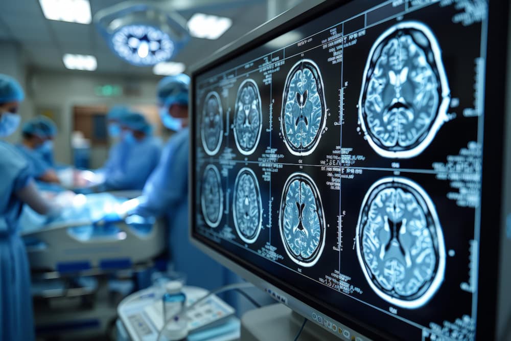

Mechanical thrombectomy is a minimally invasive endovascular procedure that physically removes a blood clot from a blocked brain artery during an acute ischemic stroke, restoring blood flow before permanent damage can spread to surrounding tissue. Delivered by specialist teams in dedicated stroke centres around the clock, it has transformed the outlook for the most severe type of ischemic stroke — a category that was, until relatively recently, beyond the reach of effective treatment.
What Is Mechanical Thrombectomy?
A stroke caused by a clot blocking one of the brain's major arteries cannot always be treated with medication alone. Mechanical thrombectomy — also known as endovascular thrombectomy — is the procedure used when a clot needs to be physically removed rather than dissolved. A specialist called an interventional neuroradiologist inserts a thin, flexible tube called a catheter through a small puncture in the wrist or groin and guides it through the body's arteries, using live X-ray imaging, all the way to the blocked vessel in the brain. A retrieval device captures the clot and withdraws it from the body, reopening the artery and restoring blood flow to the brain tissue that was starved of oxygen.
The procedure is performed entirely from within the blood vessels — no incision is made in the skull, and the brain itself is never directly operated on. It sits within the field of interventional neuroradiology, which applies catheter-based techniques to conditions affecting the brain's blood supply.
Patients sometimes encounter overlapping terms in this area that are worth understanding:
- Thrombectomy — removal of a thrombus, meaning a clot that formed directly inside a blood vessel
- Embolectomy — removal of an embolus, a clot that formed elsewhere in the body (often the heart, particularly in people with atrial fibrillation) and travelled to the brain through the bloodstream
- Thromboembolectomy — a term covering both scenarios; in practice, the same catheter technique is used regardless of where the clot originated, so these terms are frequently used interchangeably
Why Mechanical Thrombectomy Changed Stroke Treatment
Not all ischemic strokes are the same in terms of severity or treatment options. Around 87 percent of all strokes are ischemic — caused by a clot cutting off blood supply to part of the brain. Within that group, a subset known as large vessel occlusion (LVO) strokes are by far the most devastating. These occur when a clot completely blocks one of the brain's main arteries, cutting off supply to a large territory of brain tissue all at once. The result is rapid and severe: sudden paralysis down one side of the body, complete loss of speech, profound confusion, or unconsciousness.
For decades, the only acute treatment available was a clot-dissolving drug called tPA (tissue plasminogen activator). While tPA works reasonably well for smaller clots in smaller arteries, it successfully reopens a large vessel occlusion in only around 10 to 30 percent of cases — a large, dense clot in a major artery is too resistant to dissolve with medication alone. Landmark clinical trials in 2010's proved that mechanically removing the clot produced dramatically better outcomes. Patients treated with thrombectomy were approximately twice as likely to regain the ability to live independently compared with those given medication alone, and stroke guidelines across the world were updated almost immediately. The 2026 AHA/ASA guidelines have since expanded eligibility further, meaning more patients — including some who arrive at hospital many hours after their symptoms began — may now be candidates for the procedure.
How the Mechanical Thrombectomy Procedure Works
Time is the defining principle of acute stroke treatment. Every minute a major brain artery stays blocked, approximately 1.9 million brain cells die — which is the reasoning behind the phrase "time is brain" that drives the entire design of how stroke centres operate. The pathway from emergency call to clot removal is built around eliminating delay at every step.
The procedure itself is carried out in a specialist angiography suite using real-time X-ray guidance and typically takes between 30 and 90 minutes from start to finish. Most patients receive sedation so they are comfortable throughout; general anaesthesia is used in some cases depending on the patient's neurological condition. The stages are as follows.
Brain Imaging and Patient Assessment
Before any intervention takes place, the stroke team performs urgent brain scans to confirm the diagnosis, locate the blockage precisely, and determine how much brain tissue can still be saved. The critical distinction in this assessment is between tissue that has already been permanently damaged and the surrounding area that is at risk but still alive — a zone called the ischemic penumbra. Thrombectomy is racing to rescue that penumbra before it converts to irreversible infarct. The imaging used typically includes:
- CT angiography — maps the brain's arteries in detail and identifies the exact location of the occlusion
- CT perfusion or MRI diffusion imaging — distinguishes between tissue that is already dead and tissue that can still be saved if blood flow is restored quickly
- Standard brain CT — performed first to rule out bleeding in the brain, which would make the procedure unsafe
Clot Retrieval
Once the team has confirmed the patient is a candidate and imaging is complete, the procedure begins. A catheter is introduced through a small puncture in the groin or wrist and guided through the arteries toward the brain using live X-ray imaging. When the catheter reaches the clot, the specialist uses one of two main retrieval techniques — or a combination of both:
- Stent retriever thrombectomy — a small mesh device is deployed through the clot and expands to integrate it into its structure; the retriever is then pulled back, drawing the clot out with it
- Aspiration thrombectomy — a large-bore catheter is positioned directly at the face of the clot and suction is applied to draw it in; this is often used as a first-pass technique and is particularly effective for softer, fresher clots
Once the clot is removed, a final angiogram confirms that the artery has been successfully reopened and blood is flowing normally into the previously blocked territory.

Who Is a Candidate for Mechanical Thrombectomy?
Patient selection for thrombectomy is one of the most important parts of the process, and each case is assessed individually by the stroke team using a combination of clinical findings and brain imaging. The criteria have broadened significantly over the past decade. Eligibility is no longer determined primarily by the clock — it is determined by whether imaging shows that meaningful brain tissue is still salvageable, regardless of how much time has elapsed.
In general, the profile most likely to benefit from the procedure includes:
- A confirmed large vessel occlusion — one of the brain's main arteries is completely blocked by a clot
- Significant neurological symptoms indicating that a large area of the brain is at risk
- Brain imaging showing a meaningful volume of tissue that can still be rescued — the ischemic penumbra is still present
- Presentation within 24 hours of when symptoms were last known to be absent, including patients who wake up with stroke symptoms and cannot confirm an exact onset time
Several factors can affect whether the procedure is appropriate for a given patient. If scans show that most of the affected brain territory has already been permanently damaged before treatment can be given, reopening the artery is unlikely to offer benefit and could increase the risk of bleeding into the damaged tissue. Haemorrhagic stroke — where the problem is bleeding rather than a blockage — is an absolute contraindication, as thrombectomy is exclusively for ischemic strokes caused by a clot. Patients with significant pre-existing disability are considered on an individual basis.
Clinical trial data and real-world evidence consistently show that older patients, including those over 80, benefit from mechanical thrombectomy to the same degree as younger stroke patients when the right clinical and imaging criteria are met.
Mechanical Thrombectomy vs. Thrombolysis
Both treatments aim to reopen a blocked brain artery, and the question of which is appropriate — or whether both should be used — is something patients and families frequently want to understand. The two treatments work differently, suit different clinical situations, and have different limitations.
- Thrombolysis — the clot-dissolving drug treatment, most commonly tPA (alteplase) or tenecteplase — is injected into a vein and works by chemically breaking down the fibrin structure of the clot. Its main advantage is speed and availability: it can be given at any stroke-capable emergency department within minutes of diagnosis, without specialist equipment. Its limitations are a narrow treatment window of up to 4.5 hours from symptom onset for most patients, a small risk of causing bleeding in the brain, and limited effectiveness against large, dense clots.
- Mechanical thrombectomy physically removes the clot regardless of its size, composition, or resistance to medication. Recanalisation — the successful reopening of the blocked artery — is achieved in 70 to 90 percent of cases with thrombectomy, compared to around 30 to 40 percent with medication alone in large vessel occlusions. It can be used up to 24 hours after symptom onset in appropriately selected patients and is effective even when thrombolysis has already been tried and failed.
At comprehensive stroke centres, the two treatments are most often used together rather than as alternatives. Eligible patients receive thrombolysis on arrival while the specialist endovascular team assembles — a strategy known as bridging thrombolysis — and mechanical removal follows immediately after.
Thrombectomy in Other Parts of the Body
The term thrombectomy extends beyond stroke, and patients may encounter it in other clinical contexts. The underlying principle is the same — mechanically removing a dangerous blood clot — but the location, devices used, and urgency differ considerably from the cerebral setting. The most common examples are:
- Pulmonary thrombectomy (also called pulmonary embolectomy) — removal of a life-threatening clot from the pulmonary artery in the lungs, used in the most critical cases of pulmonary embolism where the clot is causing severe cardiovascular compromise
- Surgical thrombectomy — open surgical removal of a clot from a limb artery or bypass graft, used when catheter-based approaches are not suitable or have failed
- DVT thrombectomy — catheter-directed clot removal for extensive deep vein thrombosis (DVT) in the leg or pelvis, particularly in younger patients at high risk of long-term venous damage
- IVC thrombectomy — removal of clot from the inferior vena cava, the large vein that returns blood from the lower body to the heart, typically in the context of a filter complication or massive venous occlusion
None of these are the same procedure as cerebral mechanical thrombectomy. They involve different specialists, different equipment, and different clinical pathways.
Recovery After Mechanical Thrombectomy
Recovery varies considerably from one patient to the next, and it is important for patients and their families to have realistic expectations from the outset rather than a fixed timeline. Some patients experience striking neurological improvement within hours of the artery being reopened — regaining movement, speech, or vision that was lost at the onset of the stroke. This happens because restoring blood flow allows brain cells that were stunned but not yet dead to resume function relatively quickly. Others have a more gradual recovery, particularly when a significant area of brain tissue was already damaged before treatment could be given.
The factors that most influence the pace and extent of recovery are:
- How quickly the artery was reopened after the stroke began — the earlier blood flow is restored, the more tissue survives
- Which part of the brain was affected and how large the area of damage is
- The patient's age and overall health before the stroke
- The quality and consistency of rehabilitation in the weeks and months after discharge
Even when thrombectomy is technically successful, most patients need structured rehabilitation to rebuild lost function. The brain has a remarkable capacity to reorganise and adapt — a process known as neuroplasticity — and consistent, targeted therapy is the most powerful factor in maximising how much function is recovered over time.
Post-stroke rehabilitation typically involves physiotherapy to rebuild movement and balance, occupational therapy to relearn everyday activities, and speech and language therapy for patients with difficulties communicating or swallowing. The first three to six months are the most intensive period of recovery, but meaningful improvement can continue well beyond that with sustained effort.
Risks of the Procedure
Mechanical thrombectomy is a low-risk procedure relative to the severity of the condition it treats. The most important risks to be aware of are:
- Symptomatic intracranial haemorrhage — bleeding in the brain is the most serious potential complication, occurring in approximately 4 to 7 percent of patients, and can worsen neurological outcomes in affected cases
- Access site complications — bruising or minor injury at the wrist or groin puncture point, which is almost always manageable with local treatment and resolves without lasting consequences
- Vessel injury — damage to a blood vessel wall during catheter navigation is rare in experienced hands but is a recognised risk of any intracranial endovascular procedure
- Contrast reaction — a small risk of allergic reaction to the imaging dye used during the procedure, or of temporary kidney strain, particularly in patients with pre-existing kidney problems
For patients with a confirmed large vessel occlusion, the risks of not treating are far greater than the procedural risks above. Without intervention, a severe LVO stroke almost always results in permanent major disability or death.
Stroke Treatment and Mechanical Thrombectomy at Medical Park
Turkey has developed a strong network of certified comprehensive stroke centres over the past decade, and Medical Park Hospitals are among the most fully equipped in the country for around-the-clock acute stroke care. Mechanical thrombectomy is available 24 hours a day, seven days a week, in dedicated biplane angiography suites staffed by interventional radiologists and endovascular specialists with advanced cerebrovascular training. The stroke pathway at Medical Park is built around eliminating delay at every stage: when a stroke alert is activated, teams from emergency medicine, neurology, radiology, and neurosurgery mobilise simultaneously, and CT angiography with perfusion imaging is completed within minutes of a patient's arrival.
International patients come to Medical Park's Stroke Center for several distinct reasons. Some require acute stroke treatment — thrombectomy, thrombolysis, or both — when a stroke occurs while they are travelling or living in Turkey. Others arrive seeking a second medical opinion on a procedure that has already been recommended or partially performed elsewhere, such as thrombectomy, tPA treatment, surgical intervention for brain haemorrhage, or aneurysm management. Many come specifically to access post-stroke rehabilitation programmes that may not be available or affordable in their home country, including robotic physiotherapy, speech and language therapy, and neuropsychological recovery support.
For patients seeking a medical second opinion, Medical Park's multidisciplinary team reviews all existing scans, procedure reports, and clinical notes and provides a structured, independent assessment of treatment options and realistic recovery expectations. Remote consultation and care coordination are available through the international patient services team for families managing ongoing neurological follow-up from abroad — at costs that are substantially lower than equivalent private care in Western Europe or North America.





 Makaleye Dön
 E-Result
 WhatsApp